Ear cytology – a guide on what to do, what for and when

Karina Fresneda DVM, DipIACVP explains how this technique can help accurately diagnose otitis externa in cats and dogs.
Otitis externa remains one of the most frequent reasons for veterinary consultation in small animal practice, with a reported prevalence in dogs of 7.3% in primary care1.
While otitis externa can occur in cats, it is not as common as in the canine population2. In both canine and feline patients, early, accurate diagnosis and treatment is essential to prevent otitis becoming chronic or recurrent. Once established, chronic otitis can lead to fibrosis, stenosis and mineralisation of the ear canal, resulting in irreversible change.
By definition, otitis externa refers to inflammation of the epithelium lining the external ear canal, which may or may not be associated with infection3.
The condition is typically multifactorial, often arising from a combination of primary, secondary, predisposing and perpetuating factors (Panel 1)4. Its management, therefore, requires both accurate diagnosis of infection and identification of the underlying causes that allow inflammation to persist.
Panel 1. Factors behind otitis externa
Primary factors – directly cause inflammation in an otherwise healthy ear
- Atopic dermatitis
- Food allergy
- Parasites e.g. Otodectes cynotis
- Foreign bodies e.g. grass awns
- Keratinsation disorders
- Autoimmune disease
- Endocrinopathies e.g. hypothyroidism
- Neoplasia or polyps
Secondary factors – do not initiate disease but worsen it once inflammation is present
- Bacteria e.g. Staphylococcus, Streptococcus, Pseudomonas
- Yeast e.g. Malassezia pachydermatis
Predisposing factors – increase the likelihood of disease developing
- Pendulous or hairy ear canals
- Excessive moisture – swimming, frequent bathing or humid environment
- Conformation
Perpetuating factors – result from chronic inflammation and prevent resolution
- Chronic hyperplasia, fibrosis, stenosis
- Cerumen gland hyperplasia
- Biofilm formation
- Otitis media
Clinical presentation
The clinical presentation of otitis externa varies considerably depending on the underlying cause and stage of disease.
In mild or early cases, owners may simply report occasional head shaking or scratching. As inflammation progresses, the ear often becomes more painful, and changes to the pinna and canal will be visible. A full diagnostic workup begins with a detailed history and visual examination of the pinnae, followed by palpation of the canal and otoscopic inspection. Assessment should include both ears, even when disease appears unilateral.
A clinically normal ear canal is smooth, pale pink and free of discharge. When otitis is present, clinical signs may include erythema, alopecia of the pinna, excoriations
secondary to self-trauma, hyperpigmentation, crusting and, in some cases, ulceration of the ear canal.
Chronic inflammation leads to pathological changes including glandular hyperplasia, glandular dilation, epithelial hyperplasia and hyperkeratosis5. These changes progressively narrow the ear canal, impair ventilation and trap moisture, creating a self-perpetuating environment for further microbial growth.
In chronic or recurrent cases, fibrosis and mineralisation may lead to partial or complete stenosis, making topical therapy less effective and otoscopic examination difficult. Without appropriate management, this process culminates in irreversible end-stage ear disease.
Otitis may be unilateral or bilateral. Acute onset unilateral disease should prompt investigation for a foreign body, whereas a slower onset may be more indicative of polyps or neoplasia. In contrast, bilateral disease is more often secondary to underlying food allergy, atopy or other systemic conditions, including endocrinopathies.
However, due to the multifactorial nature of otitis externa, diagnosis cannot rely on clinical signs alone and cytology is an essential part of the initial workup.
Role of cytology
Cytology is a simple, quick and invaluable tool in the diagnosis and management of otitis externa. It confirms whether infection is present, helps identify the microorganisms involved and provides an objective assessment of inflammation.
In an era of increasing antimicrobial resistance, cytology provides an immediate, evidence-based method of ruling in or out microbial infection, preventing empirical treatment based solely on clinical signs. While culture and sensitivity is indicated in some cases, it can be poorly predictive of the response to topical treatment.
Cytology should ideally be carried out at first presentation and, where infection is identified, repeated until full resolution has been achieved. Topical therapy should only be stopped when cytology shows no bacteria or yeast, and no inflammatory infiltrate6. Some authors recommend weekly rechecks to help determine when this point has been reached7.
In most cases, cytology can be performed in house using a good quality light microscope and easily incorporated into existing workflows. Alternatively, samples may be sent to an external diagnostic laboratory – especially when further testing is anticipated.
Sample collection and preparation
Accurate cytology begins with an appropriate sampling technique (Panel 2). After otoscopic examination of the ear canal, debris and exudate are collected using a sterile swab. The swab should be inserted to the level where the vertical canal turns into the horizontal canal and rotated gently to collect material.
Panel 2. Top tips for ear cytology
✓ Always sample before cleaning the ear
✓ Sample both ears, even if one appears normal
✓ Roll, don’t smear to preserve morphology
✓ Look for:
- Staphylococcus spp.: pairs, fours or grape-like clusters of cocci
- Streptococcus or Enterococcus spp: chains of cocci
- Pseudomonas aeruginosa: gram-negative rods
- Malassezia pachydermatis: characteristic peanut shape
- Intracellular bacteria
✓ Repeat cytology before stopping treatment
✓ If rods are present, consider culture
Sampling both ears is recommended, even when only one appears clinically affected.
If cytology and culture are both required, the swab should first be rolled along one or two clean glass slides before being placed into a transport medium. Rolling, rather than smearing, helps preserve cell morphology. In painful ears, sedation and appropriate analgesia allow thorough assessment and easier sample collection, minimising the risk of trauma to the ear canal or tympanic membrane.
Slides should be air dried before staining. If ectoparasites are suspected, an unstained preparation can be examined under low power (4× to 10× objective) in mineral oil to look for Otodectes cynotis in dogs and cats, or Psoroptes cuniculi in rabbits. Demodex species mites may also be identified using a firm swabbing technique or superficial scraping of the pinna.
Staining is typically performed using a Romanowsky-type stain or a modified Wright’s stain. Once stained and dried, slides should be examined systematically. Examination under low power provides an overview of background material, inflammation and cellularity, while focusing on areas of interest with higher magnification (40× and 100× oil immersion) allows assessment of bacterial morphology and numbers (Figure 1).
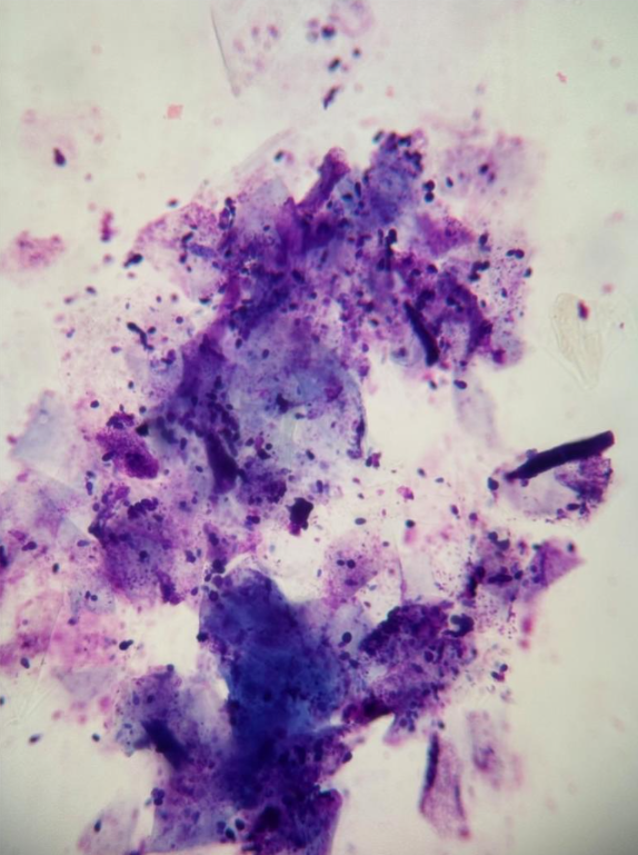
Figure 1. Ear cytology showing clusters of anucleated squamous epithelial cells with attached Malassezia species, free yeasts in the background, and small numbers of mixed bacteria, keratin and cellular debris.
The article continues beyond this point.
Read the full article https://www.vettimes.com/clinical/small-animal/ear-cytology-a-guide-on-what-to-do-what-for-and-when
Original publication: Vet Times (2025), Volume 55, Issue 50, Pages 6-8
About the author
Karina Fresneda, DVM DiplACVP, graduated from the National University of the Centre of Buenos Aires Province in 2000. She taught infectious diseases at the same university for 15 years while gaining extensive clinical and laboratory experience in pathology, cytology, and histopathology. Karina completed a three-year anatomic veterinary pathology residency at the University of California, Davis, and continues to teach courses in clinical pathology, cytology, and histopathology. For over five years, she has worked as an anatomic pathologist at NationWide Laboratories, bringing her expertise
to support our advanced diagnostic services. Learn more about our Histology and Cytology service here.