Case of Canine Pancreatitis in a four-year-old beagle x poodle

Author: Sandra Dawson, BSc BVMS FRCPath MRCVS
Clinical findings
A 4-year-old female neutered beagle x poodle presented with a 1-week history of vomiting and diarrhoea. An initial haematology profile showed a lipaemic sample with a circulating neutrophilia and monocytosis. There was a moderate increase in alkaline phosphatase (ALP) and a mild increase in alanine aminotransferase (ALT). Total bilirubin was also moderately increased. There was also a moderate hypercholesterolaemia and a marked increase in lipase (DGGR) which correlated with a similar increase in canine pancreatic lipase immunoreactivity (cPLI) consistent with pancreatitis. On ultrasound examination a suspected cystic structure was observed caudal to the right kidney with intestine and pancreas adherent on all sides. A loop of jejunum appeared to be adherent to necrotic pancreatic tissue. Biopsies were taken to rule out other conditions such as penetrating foreign body and neoplasia. The patient did not respond to attempts to stabilise the pancreatitis and was subsequently euthanised.
Pathological findings
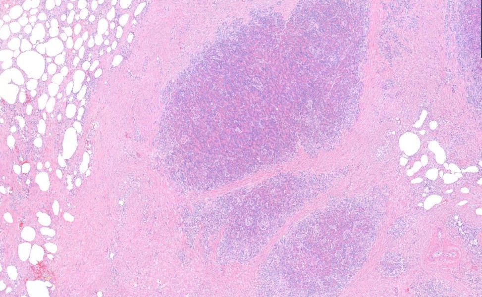
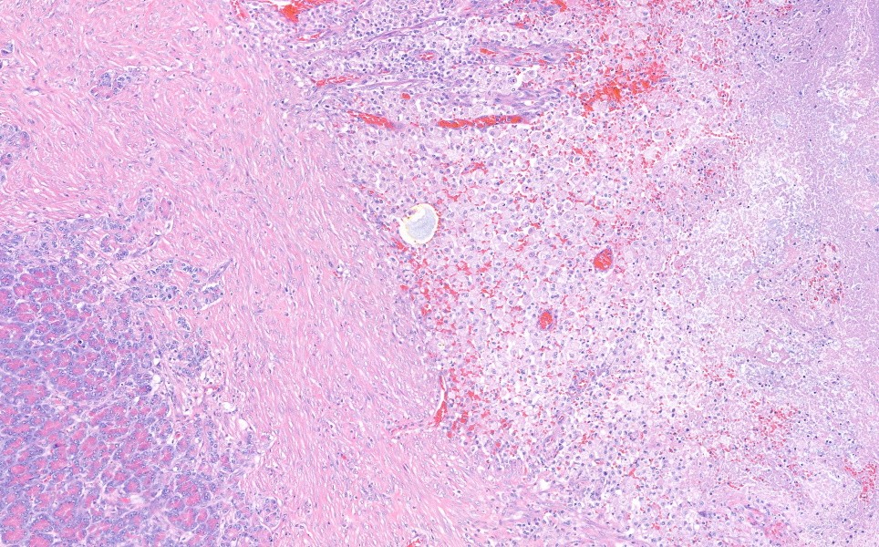
Histological sections were prepared from the pancreas and showed fibrosed peri-pancreatic fat with central foci of isolated exocrine pancreas containing scattered small pancreatic islets (fig.1). This was surrounded by extensive areas of necrotic fat and fibrous connective tissue consistent with post necrotic scarring. Fibrous tissue radiated out into the surrounding adipose tissue. There was haemorrhage with inflammatory cell infiltrates between the areas of fibrosis and necrosis (fig. 2).
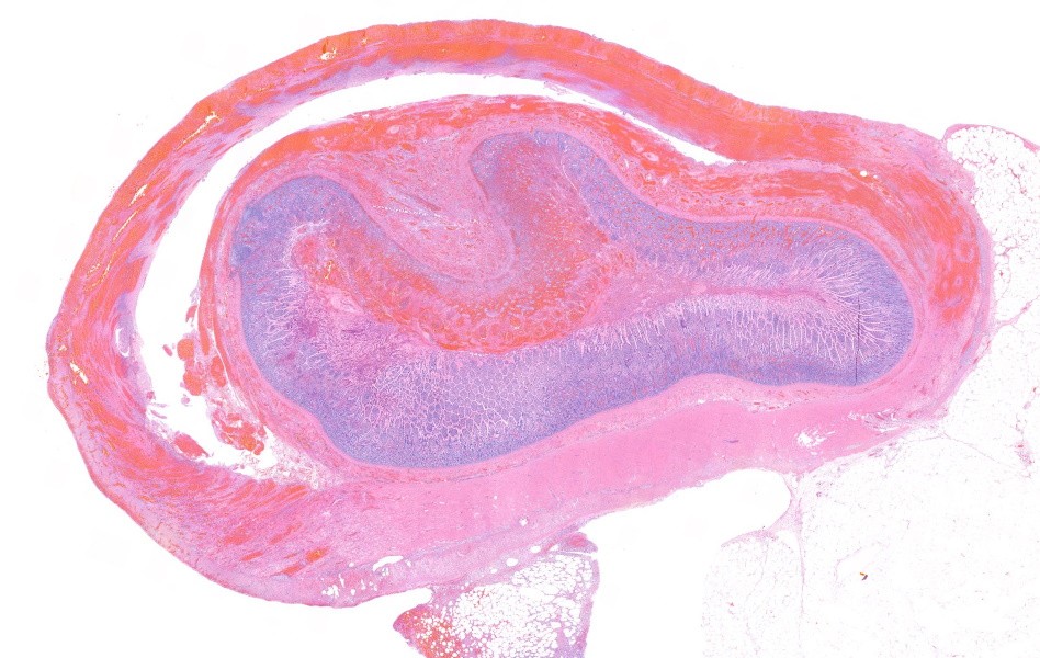
The intestine was collapsed and surrounded by a thick blood clot (fig. 3). Intestinal mucosa was congested and demonstrated areas of mucosal haemorrhage and inflammation. There was marked congestion, haemorrhage and inflammation of the submucosa and thick muscle layers. Necrotic fat, blood and inflammatory cells were adherent to the serosal surface.
Diagnosis
Acute onset pancreatitis (acute pancreatic necrosis), marked
Enteritis and transmural haemorrhage with serosal necrosis
Discussion
The histological picture is most consistent with a primary acute onset pancreatitis (acute pancreatic necrosis). This is a common disease in dogs which can present as acute or chronic disease. The condition may manifest as an acute and potentially life-threatening syndrome (as in this case) or as a chronic relapsing syndrome that may culminate in exocrine pancreatic insufficiency and diabetes mellitus. In this case the underlying cause was not evident histologically. Many cases of acute pancreatitis in dogs are believed occur after the dog has consumed a meal high in fat or some other dietary indiscretion however in reality most cases are idiopathic. Although many dogs appear to recover clinically from acute pancreatitis there may not be complete resolution of the pathological process within the pancreas. This can result in smouldering inflammation with recurrence of clinical signs, resulting in intermittent chronic relapsing pancreatitis. This case was complicated by marked necrosis spreading into surrounding mesenteric fat resulting in adhesions with intestinal wall haemorrhage with areas of inflammation due to the release of lipase from the damaged pancreas. Adhesions and inflammation have resulted in marked intestinal dysmotility and ongoing necrosis which would likely eventually result in multiorgan failure. Treatment is complex and primarily supportive and symptomatic, consisting of fluid therapy, pain relief, antiemetics, nutritional support and appetite stimulants. Continued monitoring of haematology and biochemistry parameters is also key to ensure there is a reduction in pancreatic enzymes and circulating leucocytosis as well as monitoring liver disease to look for any evidence of sudden increases in inflammatory parameters, thromboembolic disease or extrahepatic bile duct obstruction. Aspiration pneumonia is a further potential complication if vomiting persists. These cases can be challenging with little targeted therapy available. The narrative review below provides some useful clinical inflammation and discusses current, and potential future, treatment options.
References:
Lim SY, Cridge H, Twedt DC, Ohta H, Nuruki T, Steiner JM. Management of acute-onset pancreatitis in dogs: a Narrative Review. J Am Vet Med Assoc. 2024 Jun 5;262(9):1231-1240. doi: 10.2460/javma.24.02.0107. PMID: 38838711.