Interesting Hedgehog Case
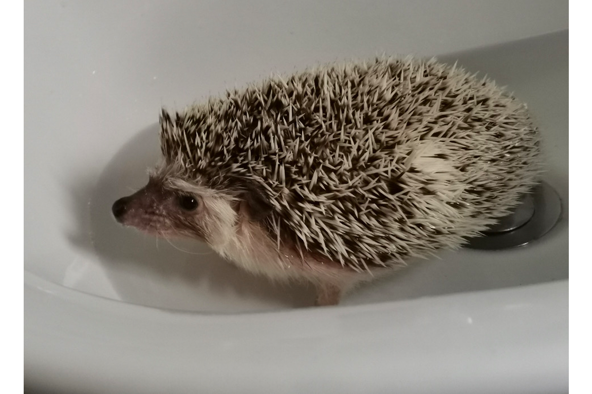
A 1-year-old African Pygmy hedgehog was presented to exotics consultant, Madonna Livingstone for ovariohysterectomy due to a bloody vaginal discharge. However, during surgery, the uterus was grossly thickened, and the internal vagina was filled with blood. Dr Livingstone sent the uterus and ovaries to the pathology team at Nationwide Laboratories where Dr Marvin Firth (anatomic pathologist) examined the sections digitally. The ovaries showed occasional primary and secondary follicles, but most of the tissue was effaced by nests and islands of neoplastic cells that were often arranged in pseudorosettes and had classic, Call-Exner bodies within them which multifocally infiltrated adjacent ovarian tissue. The cellular morphology and architecture, along with these bodies led to a diagnosis of bilateral granulosa cell tumours (GCTs). The sections of the uterus showed proliferation of the endometrial mucosa with cystic degeneration and hyperplasia of endometrial glands, occasionally forming papillary projections into the uterine lumen. Glandular structures also extended down into the muscle layers (adenomyosis). Within these layers there was also noticeable inflammation. We believe that the GCTs have led to endometrial cystic degeneration and hyperplasia with the adenomyosis and endometritis (inflammation) through production of higher levels of oestrogen. This pattern of pathology is often seen in dogs with GCTs and associated uterine changes as seen with this hedgehog. More recently, Wu et al (2021) published GCTs in 8 African Pygmy hedgehogs, which again showed the similar uterine changes observed in this case.
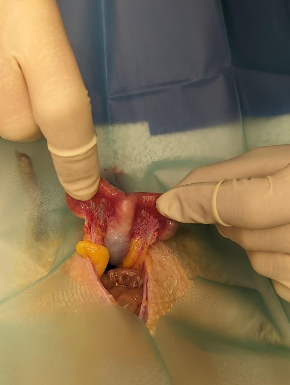
FIGURE 1. Gross enlargement of the uterine body observed by Dr Madonna Livingstone during ovariohysterectomy of a 1-year-old African Pygmy Hedgehog.
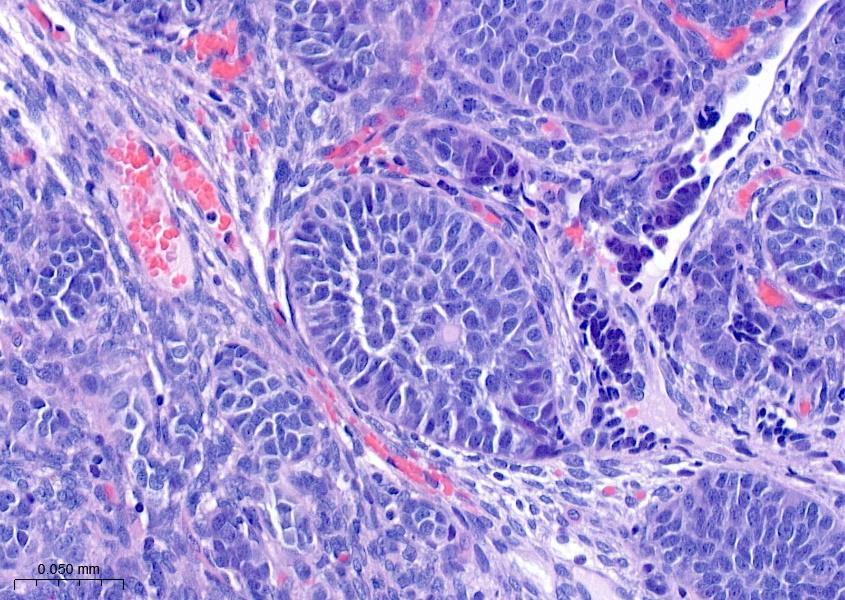
FIGURE 2: Ovary, H&E: Granulosa cell tumour (GCT) exhibiting pseudorosettes of neoplastic cells with the typical Call-Exner body, a histological characteristic in GCTs in some species.
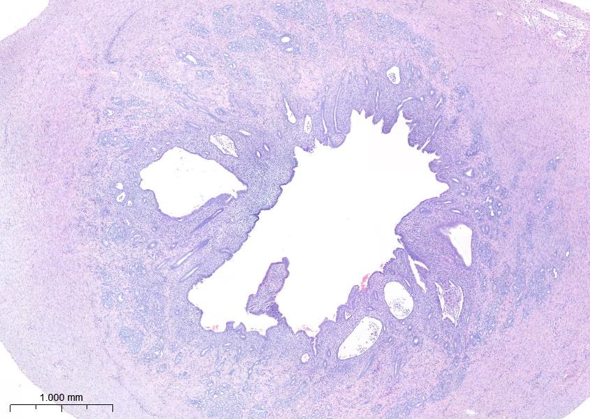
FIGURE 3: Uterus, H&E: Endometrial cystic degeneration and hyperplasia with adenomyosis and endometritis thought to have been induced by the hyperoestrogenism/hormonal effect of bilateral GCTs.
REFERENCE: Wu CC, Nakata M, Chambers JK, Uchida K, Sasaki N, Miwa Y. Granulosa cell tumor in 8 African pygmy hedgehogs (Atelerix albiventris). J Vet Med Sci. 2021 Apr 9;83(4):685-688. doi: 10.1292/jvms.20-0521. Epub 2021 Feb 17. PMID: 33597318; PMCID: PMC8111359.