Lymphangiosarcoma in a Cat
Author: Karina Fresneda DVM, Dipl. ACVP
We received a skin and subcutaneous tissue sample from the caudoventral abdomen of an 8-year-old domestic short hair cat.
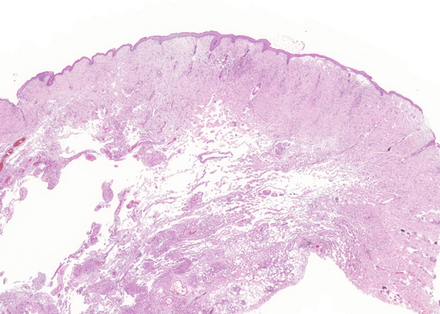
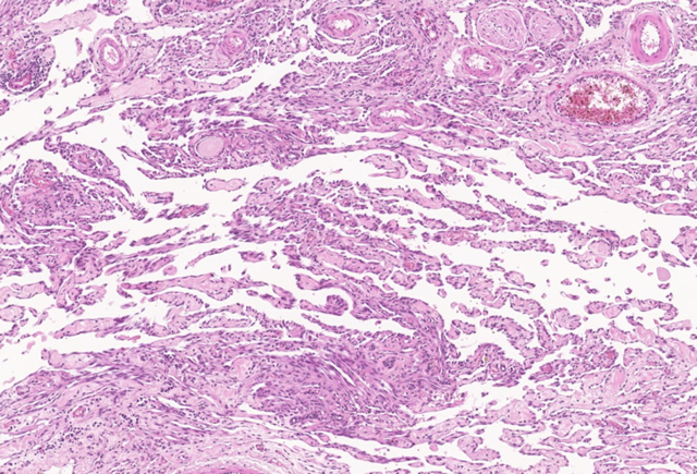
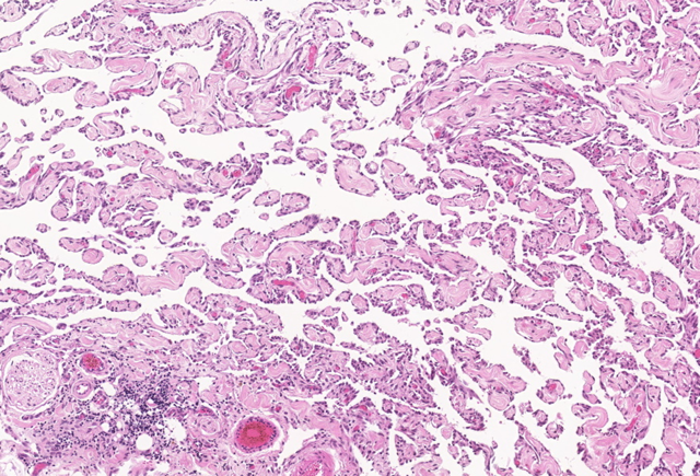
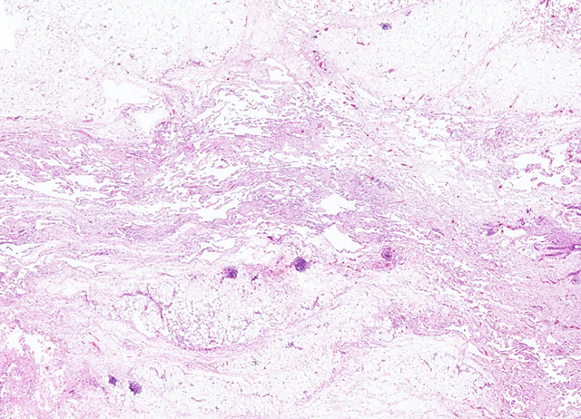
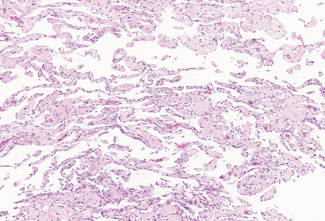
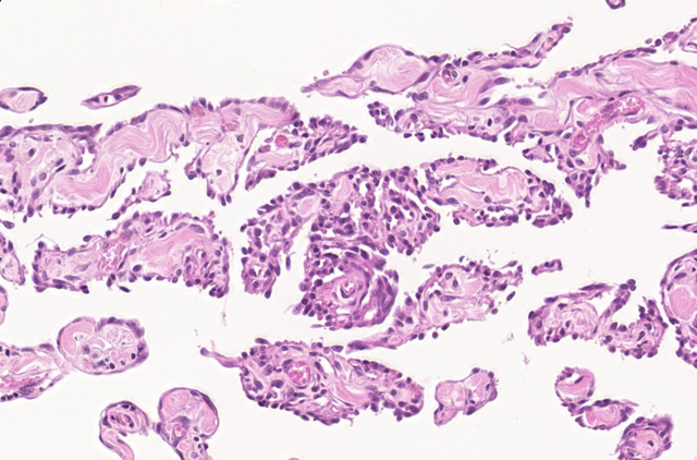
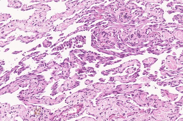
Microscopic examination revealed a non-demarcated, unencapsulated, and infiltrative proliferation of irregular vascular channels. These channels contained few to no erythrocytes and dissected between dermal collagen bundles, adipose tissue, and muscle fibers. They were lined by a single layer of round to oval endothelial cells, which occasionally protruded into the vessel lumina. The neoplastic cells exhibited basophilic cytoplasm and round to elongated nuclei, occasionally with more than one nucleolus. Mitotic figures were rare, with 0–1 per 10 high-power fields (HPFs). These histopathological features were consistent with a diagnosis of well-differentiated lymphangiosarcoma.
Lymphangiosarcoma is a rare malignant vascular neoplasm in cats, with no known breed predisposition. It typically arises within the dermis and subcutis and may occur at any anatomical site. However, it is most frequently reported in the subcutaneous tissues of the ventral thorax and abdomen, particularly the caudoventral abdominal wall. Interestingly, development of lymphangiosarcoma has also been documented in the ventrothoracic region following forelimb amputation in two feline cases. Clinically, affected areas may present with poorly circumscribed, and highly infiltrative, extensive bruising and oedema. Draining tracts that exude haemorrhagic to serosanguineous fluid are often observed and may be associated with focal, erythematous, and hyperpigmented macules. In some cases, chylous pleural and peritoneal effusions have been reported, particularly involving the cranial mediastinum and mesentery, respectively. Regional lymph node involvement may occur.
Cytology is generally not diagnostic for this tumour, and definitive diagnosis relies on biopsy and histopathological evaluation. Histologically, lymphangiosarcomas are characterised by a poorly defined proliferation of spindle-shaped endothelial cells forming vascular clefts or cavernous spaces. A key diagnostic feature distinguishing lymphangiosarcoma from hemangiosarcoma is the relative absence of erythrocytes within the abnormal vascular channels. Cytological atypia, abnormal vascular architecture, and infiltrative growth distinguish this neoplasm from normal vascular endothelial tissue. To avoid diagnostic ambiguity, the term angiosarcoma is sometimes used more broadly. Immunohistochemistry can aid in differentiation: markers such as factor VIII and CD31 confirm endothelial origin, while PROX-1 supports lymphatic lineage.
Although metastasis is considered uncommon, the prognosis is guarded due to the tumour’s locally aggressive and recurrent nature. Currently, repeated surgical excision remains the only recognised treatment option.
 |  |
 |  |
 |  |
 |
References
Bellamy E, Larsen Moberg H, Suárez-Bonnet A, Di Palma S, Murgia D, Pittaway R and Verganti S. Feline ventral abdominal wall angiosarcoma: haemangiosarcoma or lymphangiosarcoma? Clinical and pathological characteristics in nine cases. Journal of Feline Medicine and Surgery 1-9. 2024
Goldschmidt MH, Hendrick MJ. Tumors of Skin and Soft Tissues. In: Tumors in Domestic Animals, ed. Meuten DJ, 4th ed., pp.102-103. Blackwell Publishing Company, Ames, IA, 2002
Gross TL, Ihrke PJ, Walder EJ, Affolter VK: Mesenchymal Neoplasms and Other Tumors. In: Skin Diseases of the Dog and Cat, 2nd ed., pp. 748-756. Blackwell Publishing Company, Ames, IA, 2005
Hinrichs U, Puhl S, Rutteman GR, Van Der Linde-Sipman JS, And Van Den Ingh TSGAM. Lymphangiosarcomas in Cats: A Retrospective Study of 12 Cases. Veterinary Pathology Journal. 36:164–167. 1999.
Image Challenge in Veterinary Pathology: Feline Cardiovascular Diseases. Journal of veterinary pathology. Volume 55, Issue 4, July 2018
Lenard ZM, Foster SF, Tebb AJ, Russell NJ, O’Hara AJ, Lester NV. Lymphangiosarcoma in two cats. Journal of Feline Medicine and Surgery. 9, 161e167. 2007
McInnis M., White AG, Matz B, Ji-Hang Yin, Lea CH. Successful clinical remission of feline cutaneous lymphangiosarcoma with surgical resection. Vet record case reports. Volume11, Issue3. 2023.