Cyto-histopathological Correlation: Bringing The Diagnostic Picture Into Focus
By Sandra Dawson, BSc, BVMS, FRCPath, MRCVS,
Cyto-histopathological correlation refers to the comparison of cytological and histopathological diagnoses from the same lesion.1 At case level, it helps establish whether a cytology sample collected before surgical biopsy accurately reflected the disease identified on histopathology. Across multiple cases, it becomes an important quality assurance measure.
Complementary diagnostic tools
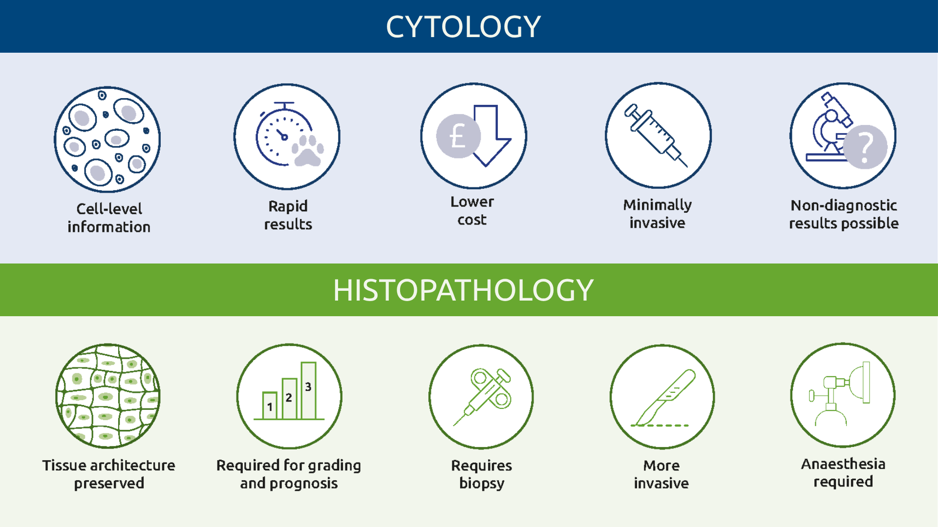
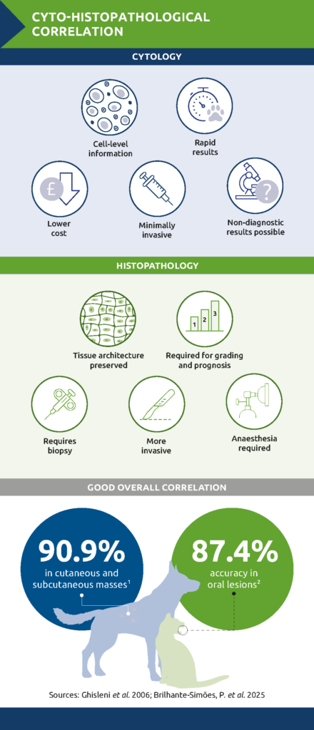
Interpreting correlation starts with understanding what each test can and cannot show. Cytology offers a rapid, minimally invasive first assessment, often ahead of any decision to proceed to surgery or biopsy, while histopathology provides the tissue-level detail needed for definitive diagnosis and grading. Cytology is often used in conjunction with histopathology and accurate interpretation of either depends on good clinical context – a recent history, relevant clinical signs and lesion-specific detail. Their respective strengths and limitations are summarised in Table 1.
| Cytology | Histopathology | |
| Invasiveness | Minimally invasive (FNA, scrape, impression or touch smears) | Requires biopsy (e.g. punch, incisional, excisional or needle core) |
| Turnaround | Rapid, sometimes in-house | Slower – requires processing, sectioning, staining |
| Relative cost | Lower | Higher |
| Tissue architecture | Not preserved | Preserved |
| Grading/prognosis | Limited information | Required for tumour grading and prognostic information |
| Main limitation | Sample may have poor cellularity or be unrepresentative of the whole lesion | More invasive; not always suitable as a first-line test |
Table 1. Cytology vs histopathology
Which lesions are well suited to cytology?
The diagnostic value of cytology varies between lesion types. Round cell tumours generally exfoliate readily and aspirates are often highly cellular; examples include lymphoma and mast cell tumours.2 Many epithelial tumours also exfoliate well, typically producing cohesive clusters or sheets of cells.
Mesenchymal lesions are less straightforward. Many, particularly benign mesenchymal lesions and soft tissue sarcomas, exfoliate poorly and may yield low-cellularity aspirates even when sampling technique is good.3 When cells are present, they are typically spindle-shaped and may be associated with extracellular matrix.
How accurate is cytology?
Published studies show good overall agreement between cytology and histopathology. Ghisleni et al. evaluated 292 non-mammary, palpable cutaneous and subcutaneous masses from dogs and cats. Cytological diagnosis agreed with histopathology in 90.9% of evaluable cases and sensitivity for diagnosing neoplasia was 89.3%. Forty-nine cases were excluded for poor cellularity, giving a retrieval rate of 83.2%.4
A more recent retrospective study of 103 oral lesions from 70 dogs and 33 cats reported a sensitivity of 84.6%, specificity of 96.0% and overall accuracy of 87.4%, after excluding samples with scant cellularity, blood contamination or formalin exposure.5
Why do cytology and histopathology sometimes disagree?
Disagreement, or discordance, does not necessarily mean either diagnosis was wrong. Sometimes, cytology and histopathology have examined different parts of the same lesion. A cytological sample can be thought of as one piece of a jigsaw – correctly interpreted but not necessarily showing the whole picture. Large or heterogeneous lesions may contain areas of inflammation, haemorrhage, necrosis and neoplasia. An aspirate taken from one area may therefore differ from a sample collected elsewhere. An impression smear from an ulcerated lesion may accurately identify surface inflammation while missing a deeper neoplasm.
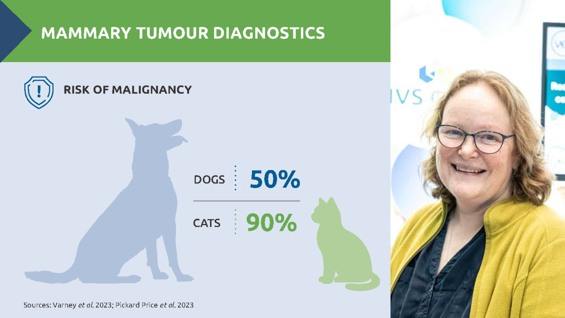
Mammary tumours provide a good example of this problem. Canine mammary tumours frequently contain a mixed population of epithelial, myoepithelial and mesenchymal elements within the same lesion. The cytological features used to distinguish benign and malignant tumours can also overlap. As a result, a cytological sample may not represent the whole lesion or reliably predict its behaviour. Cytology appears most useful for identifying simple tumour types and high-grade disease, while histopathology is necessary for definitive classification and assessment of biological behaviour.6
Timing also matters. The longer the interval between cytology and biopsy, the greater the opportunity for the lesion to progress, respond to treatment, or develop further inflammation or necrosis.
Technique and sample handling
Sampling technique is one of the most important factors influencing diagnostic quality. Poorly cellular or blood-contaminated samples can result from an unsuitable sampling site or technique – or from tissue with an inherently low exfoliative capacity. The clinician’s experience in selecting the site and preparing the slides therefore has a direct bearing on diagnostic yield. Sample handling is equally important. Crushing, thick smears, delayed drying, poor staining and exposure to formalin fumes can all obscure cellular detail.
<Box out> Improving diagnostic yield
- Select the sampling site carefully
- Sample several areas of large or heterogeneous lesions
- Avoid necrotic or haemorrhagic areas where possible
- Choose the most appropriate collection technique for the lesion
- Prepare several thin, well-prepared smears
- Air-dry slides promptly
- Label each slide with the sampling site location
- Package cytology slides separately from formalin-fixed tissue samples
- Provide all relevant history and clinical signs
Correlation as quality assurance
Across a series of cases, cyto-histopathological correlation can identify recurring problems with sampling, slide preparation, screening, interpretation or specimen handling. It may also highlight lesion types for which different sampling techniques or earlier biopsy should be considered.
Regular practice review meetings can support shared learning by confirming when the diagnostic pathway has worked well and identifying areas for improvement when findings differ. The value lies not simply in whether two reports agree, but in understanding why.
References
- Kholová, I. (2022) Cyto-Histopathological Correlations in Pathology Diagnostics. Diagnostics, 12(7) https://www.mdpi.com/journal/diagnostics/special_issues/Cyto-Histopathogical
- Cornell University College of Veterinary Medicine. Cytologic patterns, eClinpath. Available online: https://eclinpath.com/cytology/cytology-interpretation/
- Fraser, C. & Meichner, K. (2023) Skin “Lumps and Bumps” Cytology 68-77 TVP-2023-0910_Lumps_Bumps_Cytology.pdf
- Ghisleni, G. et al. (2006) Correlation between fine-needle aspiration cytology and histopathology in the evaluation of cutaneous and subcutaneous masses from dogs and cats. Veterinary Clinical Pathology. 2006;35(1):24–30. DOI: 10.1111/j.1939-165X.2006.tb00084.x
- Brilhante-Simões, P. et al. (2025) Association between cytological and histopathological diagnoses of neoplastic and non-neoplastic lesions in oral cavity from dogs and cats: an observational retrospective study of 103 cases. Veterinary Sciences. 12(2):75. DOI: 10.3390/vetsci12020075
- Dolka, I. et al. (2018) Diagnostic efficacy of smear cytology and Robinson’s cytological grading of canine mammary tumors with respect to histopathology, cytomorphometry, metastases and overall survival. PLoS ONE, 13(1), e0191595. DOI:10.1371/journal.pone.0191595
About the author:
Sandra Dawson graduated from Aberdeen and Glasgow universities with degrees in agriculture and veterinary medicine. After working in mixed practice, she completed a residency in veterinary pathology at Edinburgh University, where she also lectured in reproductive pathology.Following five years in industry, during which she gained her FRCPath, Sandra joined NationWide Laboratories. In 2011, she was appointed to the Royal College of Pathologists board of examiners in the speciality of small domestic animals. Sandra specialises in histopathology, working with surgical biopsies and fine needle aspirates across a wide range of species. Original publication: The Veterinary Edge, Issue 67, September 2026, p46/47