Case of Intraocular Heterotopic Bone Formation in a Pet Guinea Pig (Cavia porcellus)
Author: Sofia Clara Sacco DVM, PhD., MRCVS
Case presentation
A four-year-old female guinea pig (Cavia porcellus) was examined at a veterinary clinic for right ocular abnormalities. Enucleation of the right eye was performed due to suspected glaucoma and lens luxation. The surgery was complex, and intraoperative damage to the globe occurred. The enucleated eye was subsequently submitted to NationWide Laboratories for histopathological evaluation. During macroscopic examination and trimming, a firm area consistent with hard tissue was noted, and decalcification was required to allow further histological processing.
Histological findings
The globe was collapsed. Expanding from the ciliary body and extending between the corneoscleral layer and the uvea, there was a marked proliferation of well-organised lamellar bone with multiple central cavities, occasionally containing erythrocytes and a few lymphocytes and plasma cells. The bone was multifocally and partially surrounded by fibrous connective tissue containing reactive fibroblasts. No cellular atypia or mitotic figures were observed in these sections. The filtration angle was occluded by the previously described bone. The retina was detached (artefactual).
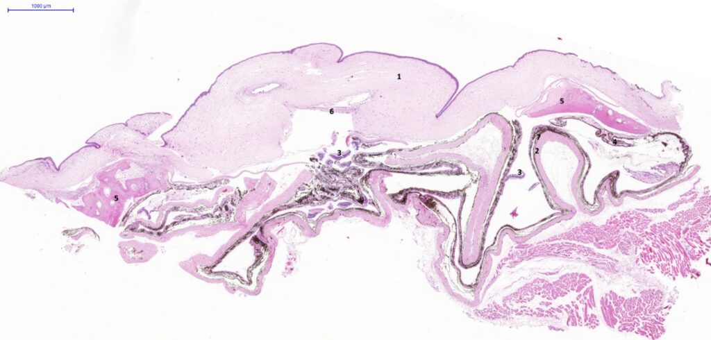
Fig. 1. Eye, guinea pig. Section of a collapsed eyeball. 1. Cornea; 2. Sclera; 3. Detached retina; 4. Ciliary processes; 5. Heterotopic bone formation; 6. Fragment of lens capsule; 7. Choroid. H&E stain. Scale bar = 1000 µm.
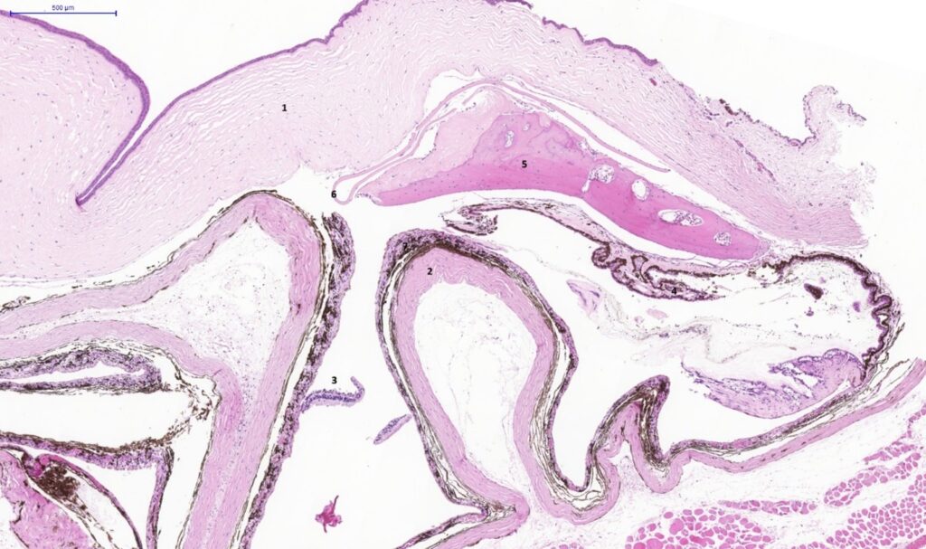
Fig. 2. Eye, guinea pig. Section of a collapsed eyeball. 1. Cornea; 2. Sclera; 3. Detached retina; 4. Ciliary processes; 5. Heterotopic bone formation; 6. Fragment of lens capsule; 7. Choroid. H&E stain. Scale bar = 500 µm.
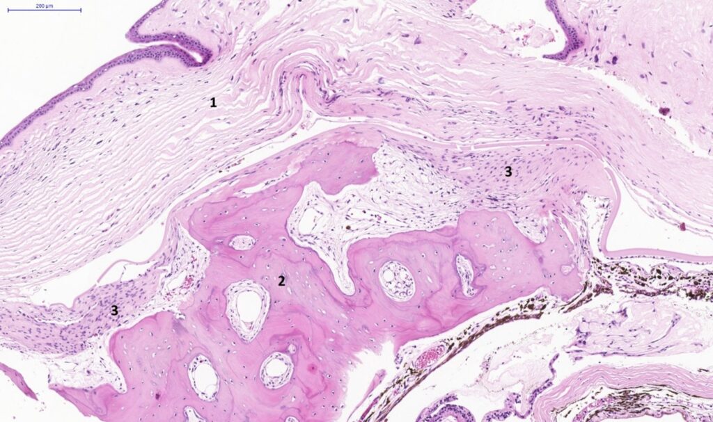
Fig. 3. Eye, guinea pig. Section of a collapsed eyeball. 1. Cornea; 2. Heterotopic bone formation; 3. fibrous connective tissue. H&E stain. Scale bar = 200 µm.
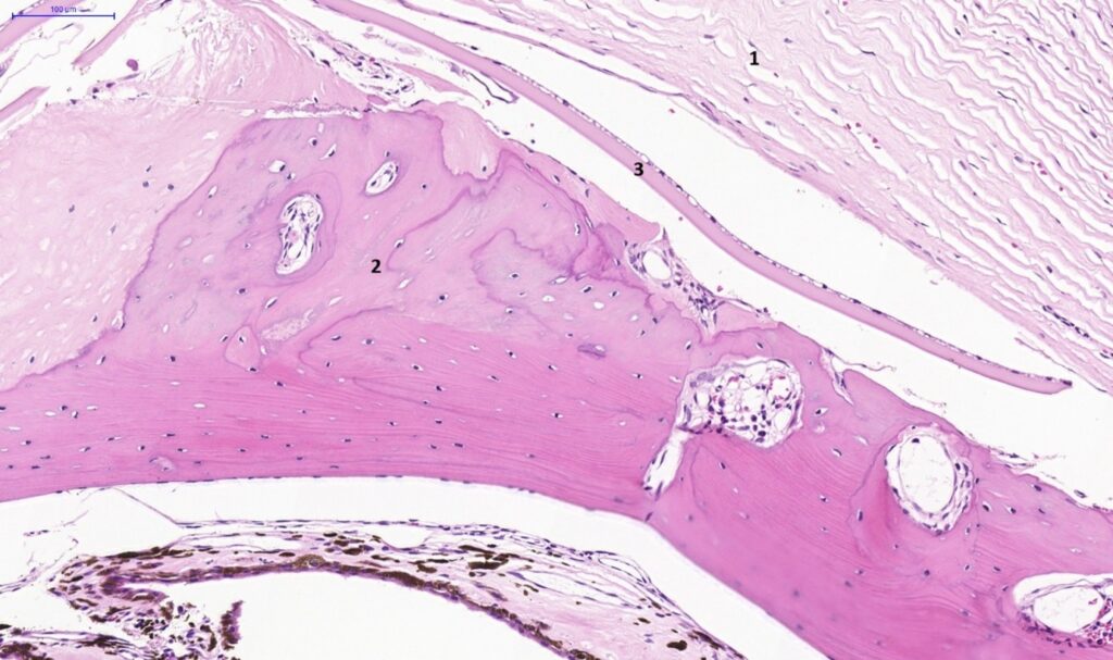
Fig. 4. Eye, guinea pig. Section of a collapsed eyeball. 1. Cornea; 2. Heterotopic bone formation; 3. Fragment of lens capsule. H&E stain. Scale bar = 100 µm.
Interpretation
Based on the microscopic findings, a diagnosis of heterotopic bone formation involving the ciliary body and extending between the corneoscleral layer and the uvea of the right eye was made.
Discussion
Microscopically, this eye exhibited features consistent with heterotopic bone formation (also referred to as an osseous choristoma), accompanied by occlusion of the filtration angle. This finding may be associated with secondary glaucoma. Heterotopic bone formation is a sporadic and usually incidental finding in the eyes of guinea pigs. It has been reported in animals of both sexes and across all age groups, occurring either unilaterally or bilaterally. Most affected animals remain asymptomatic; however, secondary open-angle glaucoma may develop if the iridocorneal filtration angle becomes obstructed, with chronic exposure keratitis described as the most notable sequela. Conversely, some studies have reported no significant elevation of intraocular pressure in affected eyes.
The precise aetiology remains unclear. While intraocular bone formation can result from metaplastic or neoplastic processes, most reported cases lack evidence of previous ocular trauma or disease. Because of their slow-growing and well-differentiated nature, these lesions are generally regarded as choristomas, representing histologically normal tissue arising ectopically as a consequence of disordered embryogenesis.
A potential contributory factor may be the ability of the ciliary epithelium to concentrate ascorbic acid within the aqueous humour, as ascorbic acid promotes trabecular bone formation through modulation of bone matrix gene expression in osteoblasts. Osseous metaplasia and mineralisation are also commonly observed in other organs of guinea pigs, particularly within the lungs.
REFERENCES
Goto, S., Zhu, Q., Jensen, K., Torres, J. A., & Wildsoet, C. F. (2021). Ocular heterotopic bone formation in a guinea pig: A case report with 7-month follow-up using advanced ophthalmic imaging technology. Clinical Case Reports, 9(4), e4076.
Donnelly, T. M., Brown, C., & Donnelly, T. M. (2002). Heterotopic bone in the eyes of a guinea pig: osseous choristoma of the ciliary body. Lab Animal, 31(7), 23–25.