Making Sense of Mammary Masses
By Sandra Dawson, BSc, BVMS, FRCPath, MRCVS
Mammary masses are common in small animal practice, particularly in older dogs and cats. However, they encompass a diverse range of lesions, from benign hyperplasia and cystic change to highly aggressive carcinomas. And importantly, not every lump in the mammary region actually originates from mammary tissue.
Skin and subcutaneous tumours, including mast cell tumours, enlarged lymph nodes and inflammatory lesions can all occur in this area. This makes a structured diagnostic approach important, both to establish what the lesion is and to provide the information needed to plan treatment.
Understanding the risk
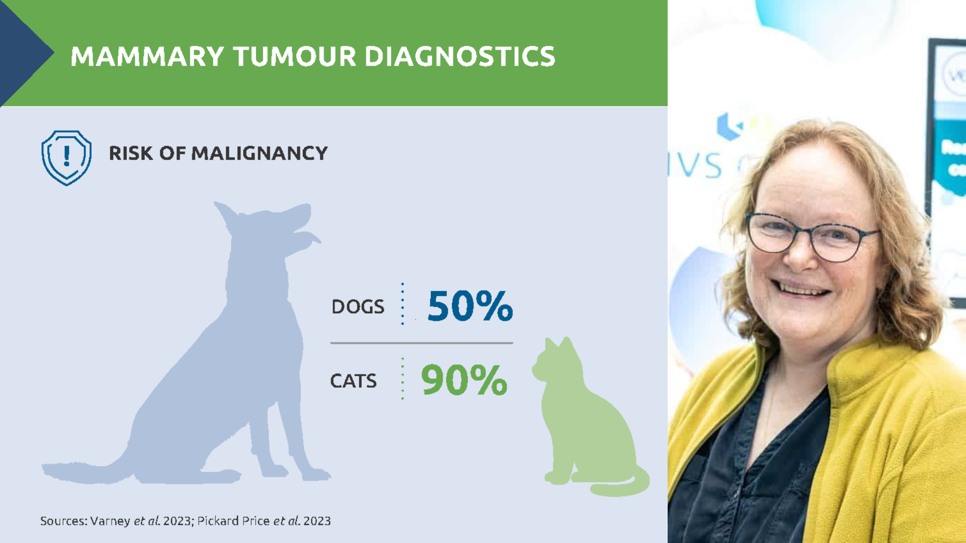
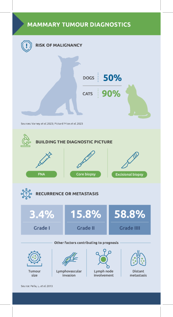
Increasing age is one of the clearest risk factors for mammary neoplasia. There are also important differences between species. Around 50% of canine mammary tumours are malignant,1 compared with approximately 90% in cats.2 In addition, feline mammary tumours also tend to behave more aggressively, with a median survival time of 8 to 12 months post diagnosis.
Hormonal influences are also important. Mammary tissue undergoes considerable physiological change during the reproductive cycle and both endogenous hormones and the administration of exogenous progestogens can cause marked mammary hyperplasia; prolonged mammary hyperplasia has been associated with malignant transformation.
Conversely, early neutering is generally associated with a reduced risk of mammary neoplasia,3 although the size of this protective effect remains uncertain and decisions around the optimal age for neutering need to take account of wider health considerations.4
Choosing the right diagnostic approach
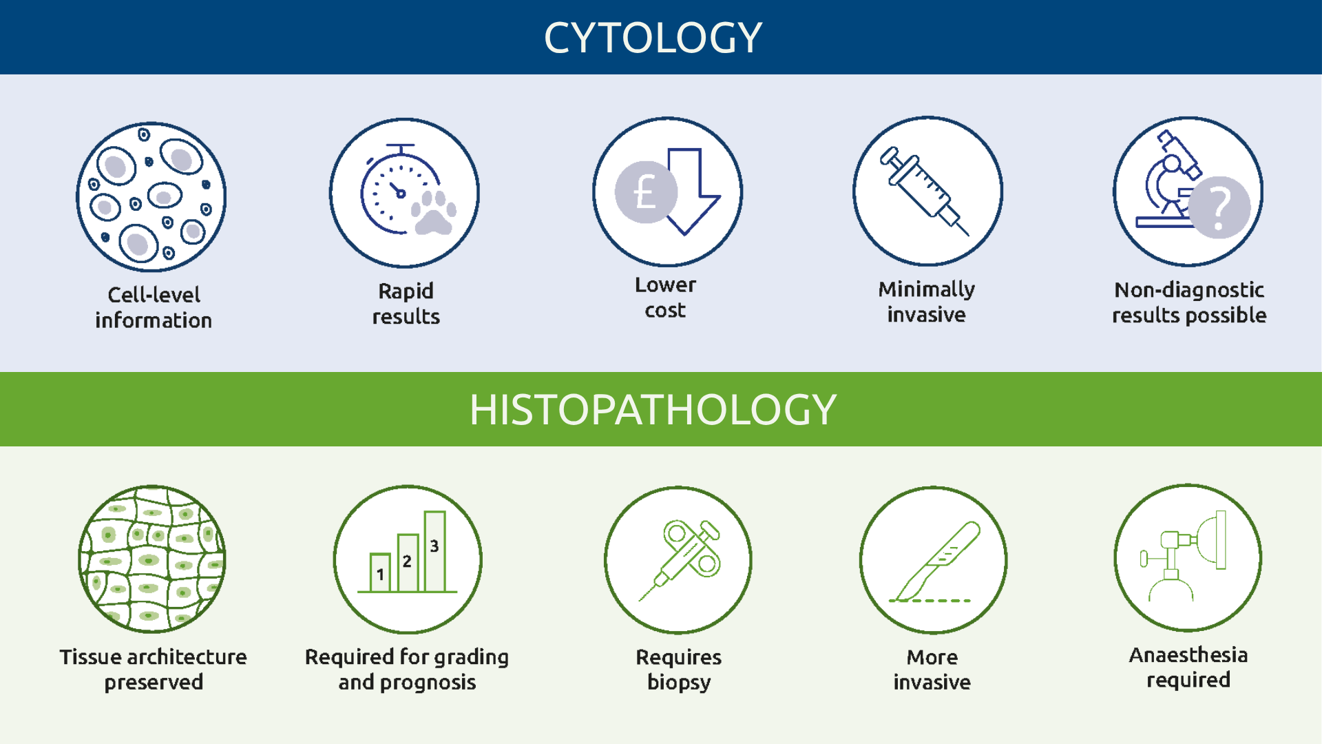
When a mammary-region mass is identified, the first question is how best to sample it. Fine needle aspiration (FNA) can be useful in some circumstances. An aspirate containing mature adipocytes, for example, may support a diagnosis of lipoma while identification of granular round cells may reveal a mast cell tumour and substantially alter the surgical plan.
For primary mammary lesions cytology tends to be less useful. Mammary tumours are often heterogeneous and cytological appearance does not always correlate with biological behaviour. Benign tumours can display cellular atypia, while well-differentiated carcinomas may contain relatively uniform cells. Histopathology therefore remains essential for definitive classification of most mammary masses, allowing assessment of tissue architecture, invasiveness, surgical margins and other features associated with malignancy.
One useful way to view this is as a diagnostic jigsaw: an FNA provides a small number of pieces, a core biopsy provides more, while examination of a larger representative tissue sample allows the pathologist to see much more of the complete picture. Excisional biopsy is the ideal sample as it allows the pathologist to examine the margins and assess whether the lesion is well demarcated or invasive.
When mammary disease does not look like neoplasia
Not every mammary neoplasm presents as a discrete nodule. Anaplastic mammary carcinoma may present with marked inflammation, sometimes described clinically as inflammatory carcinoma. Rather than forming a discrete mass, tumour infiltration and obstruction of dermal lymphatics may cause marked inflammation, oedema and ulceration, closely resembling severe mastitis.
Histologically, neoplastic cells may be scattered and difficult to identify within extensive inflammation and fibrosis. However, where this presentation is suspected, a core or punch biopsy may be preferable to a large wedge biopsy because the affected tissue can heal poorly.
Another striking differential, particularly in young cats, is fibroadenomatous or fibroepithelial hyperplasia. This can cause rapid and extensive enlargement of mammary tissue and may occur in young hormonally active or pregnant queens, as well as males or females exposed to progestogens. Despite its dramatic appearance, it is a hyperplastic rather than neoplastic process and should regress once the hormonal stimulus is removed.
Help the pathologist complete the picture
The information submitted with the tissue can be just as important as the sample itself. Essential details include:
- Age, breed and sex
- Exact location and size of the lesion
- Number of mammary masses
- Reproductive status
- Age at neutering
- In entire animals, timing of the most recent oestrus
Where several masses are removed, they should be individually identified rather than placed together without labelling. This allows histological findings, margins and prognosis to be related back to the correct lesion. For larger specimens such as mammary strips adequate formalin penetration is critical (Box 1)
Box 1: Preparing a mammary strip for histopathology
- Fix the specimen in 10% neutral buffered formalin, ideally 10 parts fixative to 1 part tissue
- Allow 24-48 hours for fixation
- Make partial incisions from the skin surface approximately 1 cm apart to improve formalin penetration
- Do not cut through the deep surface as this can compromise assessment of the deep surgical margin
- Once fixed, remove the specimen using PPE in a well-ventilated area
- Wrap in wet paper towel to prevent drying during transport
- Place in a sealed bag, then double bag and send with a fully completed submission form
From diagnosis to prognosis
Histopathology also provides important prognostic information. Mammary carcinomas can be graded according to features including tubule formation, nuclear pleomorphism and mitotic activity, with increasing grade associated with more aggressive biological behaviour.
In dogs, a prospective study using a canine-adapted histological grading system found recurrence or metastasis in 3.4% of grade I tumours, compared with 15.8% of grade II and 58.8% of grade III tumours.5
Tumour size is another important prognostic factor. In dogs, the conventional staging system categorises mammary tumours as less than 3 cm, 3–5 cm or greater than 5 cm, with tumours greater than 3 cm in diameter associated with a poorer prognosis. However, recent evidence suggests that important prognostic differences may exist even among tumours smaller than 3 cm.6
In cats, tumours measuring 2 cm or more have been associated with poorer survival.7 Other adverse findings include lymphovascular invasion, regional lymph node involvement and distant metastatic disease.
For clinicians, the key is to combine these findings rather than relying on any single feature. Tumour type, grade, size, invasion and clinical stage together provide a much more meaningful assessment of likely biological behaviour and help determine the most appropriate next step.
Mammary pathology can be complex, but good communication between clinician and pathologist makes interpretation considerably easier. A representative sample (ideally excisional biopsy), accurate clinical history and careful specimen identification all help the pathologist provide the most clinically useful diagnosis and prognostic information.
References
- Varney, D, et al. (2023) Epidemiology of mammary tumours in bitches under veterinary care in the UK in 2016. Vet Rec. e3054. https://doi.org/10.1002/vetr.3054
- Pickard Price, P., Stell, A., O’Neill, D., Church, D. and Brodbelt, D. (2023) Epidemiology and risk factors for mammary tumours in female cats. J Small Anim Pract, 64: 313-320. https://doi.org/10.1111/jsap.13598
- Guirguis, P., & Beggs, D. S. (2025) Systematic Review: Does Pre-Pubertal Spaying Reduce the Risk of Canine Mammary Tumours? Animals, 15(3), 436. https://doi.org/10.3390/ani15030436
- Stavisky, J. & White, C. (2022) Does spaying at a younger age reduce dogs’ risk of developing mammary tumours?. Veterinary Record, 190: 123-125. https://doi.org/10.1002/vetr.1455
- Peña, L. et al. (2013) Prognostic Value of Histological Grading in Noninflammatory Canine Mammary Carcinomas in a Prospective Study With Two-Year Follow-Up: Relationship With Clinical and Histological Characteristics. Veterinary Pathology. 50(1):94-105. doi:10.1177/0300985812447830
- Vieira, T.C. et al. (2025) Small canine mammary tumours have different prognostic factors: A study of 3,470 tumours. J Comp Pathol. 219:6-10. doi: 10.1016/j.jcpa.2025.02.004
- Dagher, E. et al. (2019) Feline Invasive Mammary Carcinomas: Prognostic Value of Histological Grading. Veterinary Pathology. 56(5):660-670. doi:10.1177/0300985819846870
- About the author:
Sandra Dawson graduated from Aberdeen and Glasgow universities with degrees in agriculture and veterinary medicine. After working in mixed practice, she completed a residency in veterinary pathology at Edinburgh University, where she also lectured in reproductive pathology.Following five years in industry, during which she gained her FRCPath, Sandra joined NationWide Laboratories. In 2011, she was appointed to the Royal College of Pathologists board of examiners in the speciality of small domestic animals. Sandra specialises in histopathology, working with surgical biopsies and fine needle aspirates across a wide range of species.
Original publication: The Veterinary Edge, Issue 68, October 2026, p42/43