The Spindle Cell Dilemma and the Importance of the Follow-up Biopsy

Author: Sandra Dawson
Cytological evaluation of fine needle aspirates can provide critical information to veterinary practitioners, for example in the detection of mast cell neoplasia. However, in some cases the cytological evaluation does not provide a definitive diagnosis, but it can give us some useful information and help us to determine the next step. In these instances, it is important to have a differential list in mind and to communicate this to the client.
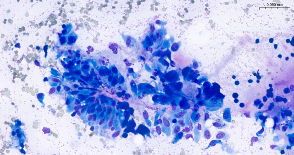
Inga is a 2-year-old female neutered golden retriever who presented to her local practice with a subcutaneous mass on her left lower abdomen which had been present for approximately 3 weeks. Initial cytological evaluation revealed some blood with low numbers of small nongranular mononuclear round cells and some large spindle cells. Spindle cells were loosely aggregated with oval nuclei containing single prominent nucleoli and stippled chromatin. Cells had abundant fibrillar or fusiform cytoplasm. There was some anisocytosis and rare binucleate cells were present. Convincing infectious organisms and other nucleated cells were not evident (Figure 1).
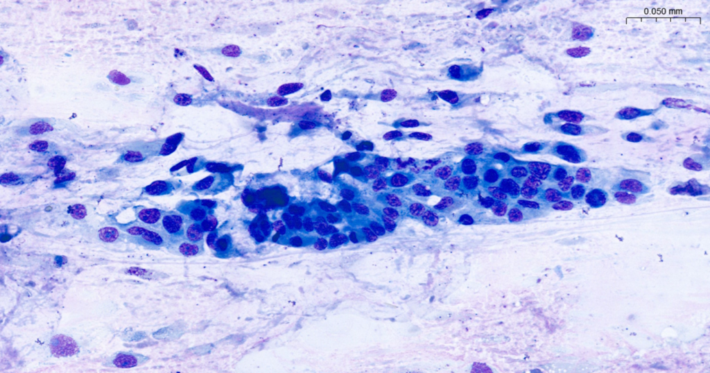
Around the same time Connie, a 13-year-old female neutered golden retriever also presented at her local practice with a small, firm cutaneous lump on her dorsal neck. Fine needle aspirates showed high cellularity. There were aggregates of spindle cells that exhibited mild anisocytosis and anisokaryosis, a mixed inflammatory process characterized by the presence of macrophages, neutrophils, necrotic cellular debris, and a small amount of blood. Spindle cells exhibited round to oval nuclei with prominent nucleoli, condensed, coarse chromatin, and abundant cytoplasm with occasional vacuolation. No binucleated cells were seen (Figure 2).
The images in each case are very similar and, in both cases, a reactive or neoplastic process was indicated, and the practitioner was advised to proceed to surgical excision with histological examination to try to reach a definitive diagnosis.
The biopsy from the younger dog consisted of skin and tissue measuring 32 mm with a firm mass measuring 19 mm. Figure 3 shows the histological presentation.
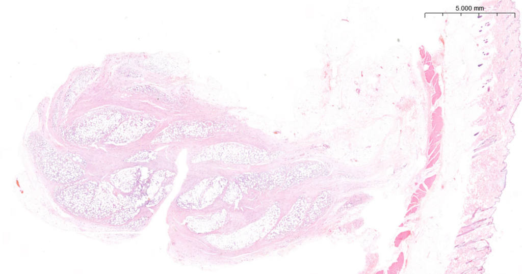
This consists of mature adipose tissue subdivided by thick transecting fibrous septae composed of large, interweaving reactive fibrocytes. There is some interstitial inflammation involving vesiculated macrophages and low numbers of lymphocytes or plasma cells. There are no convincing foci of infection or embedded foreign material. The lesion is relatively well demarcated but not encapsulated. Inflammatory cells and fibrocytes extend to the borders of the sections (Figure 4).

Convincing malignancy is not evident. The diagnosis was considered to be nodular fibrosing panniculitis which is a reactive fibroproliferative lesion. It is believed to be the result of local injury. Lesions are asymptomatic and pain is not noted. Nodules can be surgically excised or monitored to ensure that they gradually reduce in size.
Connie’s biopsy consisted of skin and tissue measuring 68 mm with a firm, raised mass measuring 28mm. The histological appearance can be seen in Figure 5 and shows a central dermal, poorly demarcated mass extending into the subcutis.
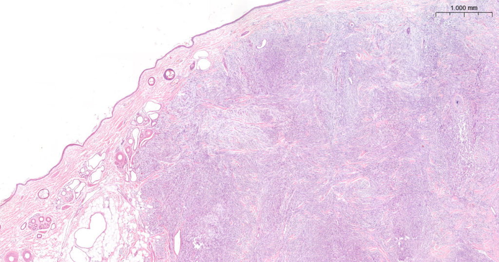
There is central necrosis and oedema. At the periphery there is haphazard proliferation of neoplastic spindle cells. Superficially these are well-differentiated, forming short interweaving fascicles and palisades. In the deeper tissue the cells become dense and pleomorphic (Figure 6).
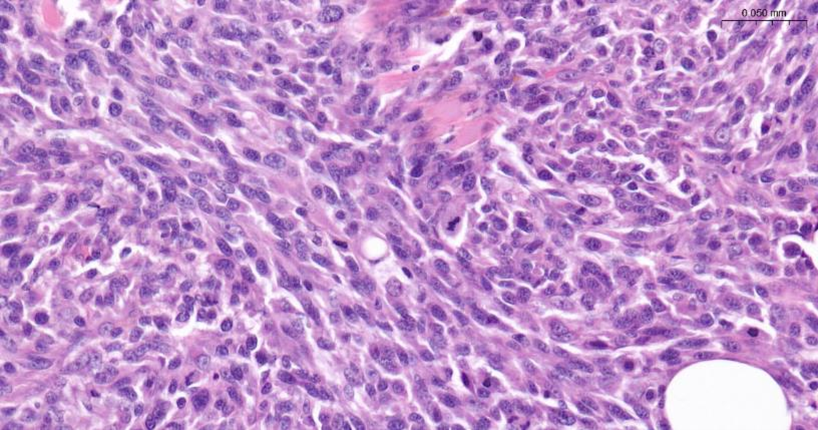
They have oval nuclei with prominent nucleoli and clumped chromatin patterns. They have fusiform cytoplasm, often with indistinct borders. There is moderate anisocytosis with scattered multinucleate cells and typically 18 mitotic figures per ten high-power fields (X 400). The lesion is not encapsulated. It is surrounded by adipose tissue which contains a few lymphocytes, plasma cells and macrophages. Margins measure 7-15 mm laterally and 3-7 mm at the deep border. The diagnosis was an intermediate-grade canine soft tissue mesenchymal tumour (differentials include fibrosarcoma, peripheral nerve sheath tumour and perivascular wall tumour). This class of tumours all behave in the same way regardless of the exact cell of origin. They are locally infiltrative and frequently recur following surgery, despite the appearance of complete excision, due to difficulty in achieving adequate excision margins at awkward surgical sites and the ability of these tumours to dissect between fascial planes. Metastasis however is uncommon or develops late in the course of the disease, usually after repeated excisions, via haematogenous rather than lymphatic spread. This particular lesion does appear to have been excised, however caution was advised, with close clinical monitoring to check for local regrowth.
The striking feature of these cases is the similarity in their cytology presentation, with both fine needle aspirates showing a population of somewhat pleomorphic spindle cells. In both cases a neoplastic lesion was a significant concern. Age may be considered an important factor in these cases, with the younger dog perhaps more likely to develop a reactive lesion over a neoplastic one and this was confirmed in the surgical biopsies.
These cases highlight the importance of following up fine needle aspirates with histological examination of the surgically excised mass. This means that the diagnosis can be confirmed and, if appropriate, any confirmed neoplasm can be graded. Additionally, the margins can be confirmed. All of this information can be used to plan for any revision surgery or adjuvant therapy.
Submission of a full signalment and history is fundamental. This allows the pathologist to formulate an appropriate differential list which can provide the clinician with key information to discuss with the owners and allow for appropriate forward planning.
About NationWide Laboratories
NationWide Laboratories is a leading provider of veterinary diagnostic services in the UK and worldwide. With over 40 years of experience, they offer a comprehensive range of tests and expert interpretation to help vets make informed decisions. Their teams can assist clients in selecting the right tests for companion, exotic, and farm animals, as well as support custom projects. As part of NVS Group, NationWide Laboratories is committed to upholding the highest standards of quality and reliability in their services.