The benefits of digital cytology and histology working together

Credit: Kerry Freel, Roberio Gomes Olinda
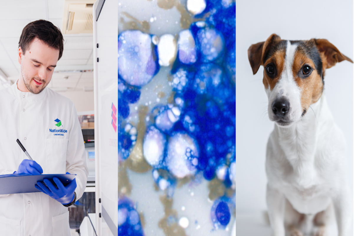
A 13-year-old Jack Russell terrier presented with a fast-growing, non-painful dermal swelling on the hind leg that had been present for 3 weeks. Both fine needle aspirates for cytology and a further excisional biopsy were taken for analysis.
The FNA preparation was sent to our laboratory in Poulton and then scanned using our 3DHISTECH digital scanner. Images such as the one below were then analysed by our experienced internal and international team, including cytologist, Roberio Gomes Olinda.

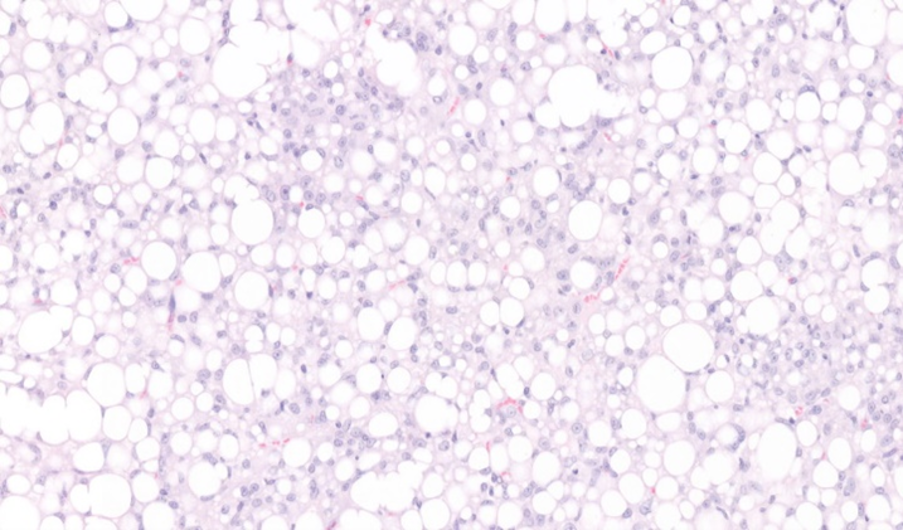
To enable a definitive diagnosis, determine a prognosis and ascertain if surgical margins were achieved, an excisional biopsy was recommended in the initial report. The presenting veterinary surgeon was then able to undertake the surgery and submitted the mass to the same laboratory for histopathological preparation and evaluation. Once again, the digital scanner was able to scan prepared haematoxylin and eosin-stained sections and experienced histopathologist, Kerry Freel was able to confirm the diagnosis.


Liposarcomas are malignant, locally invasive neoplasms of adipocytes. The tumours consist of malignant lipoblasts and mesenchymal tissue and are soft-tissue neoplasms. These tumours are primarily confined to the subcutaneous tissue of the skin, but primary tumours are also seen in the tongue, intestines, bone, spleen and liver. Metastases have been reported in some cases (to the lung, liver and bone) but these are considered rare occurrences. Excision with clear margins is often curative and, in this case, clear margins have been achieved around the mass.
The diagnosis was confirmed as a liposarcoma on the histopathology, and surgical margins reported that the mass was fully excised. Digital pathology allows for accurate measurements of surgical margins of masses that are removed surgically as well as comparison of both cytological and histopathological findings.
About NationWide Laboratories
NationWide Laboratories is committed to making a positive impact on animal health by offering innovative products, technology and laboratory services. We offer friendly advice and rapid, reliable results to help fulfil your diagnostic and therapeutic objectives, and our expert teams can assist you in making decisions on relevant testing for companion, exotic and farm animals. We offer full interpretation across a range of testing areas, including biochemistry, haematology, cytology, histopathology, endocrinology, microbiology and more.
NationWide Laboratories is focused on keeping our service modern and relevant to the veterinary field. We have added a new, extra-fast and super-efficient slide digitalisation system into the workflow, which means improved turnaround times and better first-time results for you. We are also able to receive images from practices and offer a slide scanning service.
NationWide Laboratories is customer-focused, passionate about animal welfare and always advancing! Our teams have the depth of knowledge and experience to help you help your clients.


