Superficial Pustular Dermatophytosis of the Nasal Skin in a Jack Russell Terrier
Author: Sofia Clara Sacco DVM, PhD.
Case presentation
A 10-year-old neutered male Jack Russell Terrier was presented to a veterinary clinic with a history of an ulcerative skin lesion on the nasal skin. The lesion was now scabbing and scaling but did not involve the nasal planum. The lesion had been present for 3 months and had been treated with steroids and antibiotics, with some response. In addition, the patient showed mild periorbital alopecia. Three 4 mm skin punch biopsies were taken from the nasal skin, and the samples were submitted to NationWide Laboratories for histopathological evaluation.
Histological findings
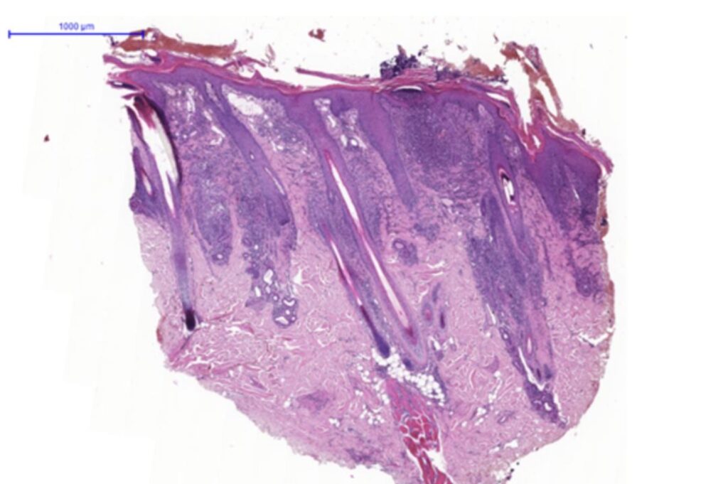
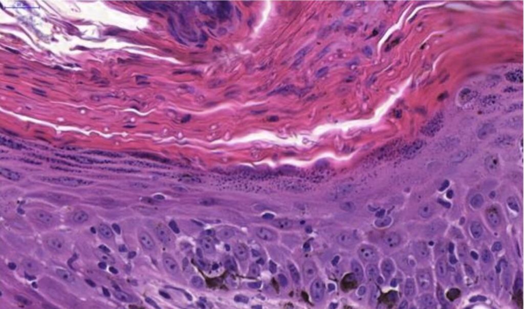
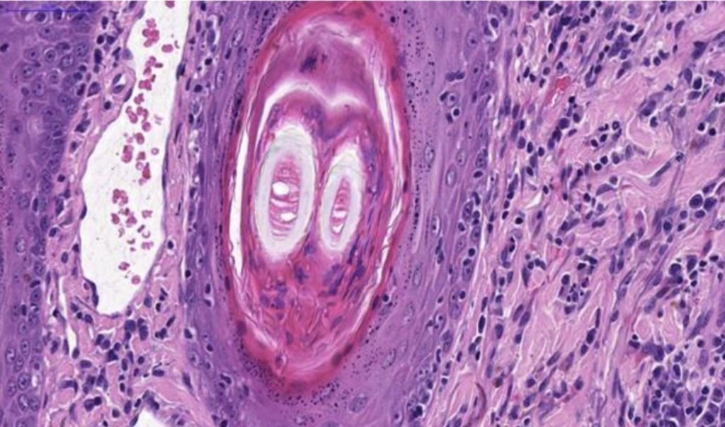
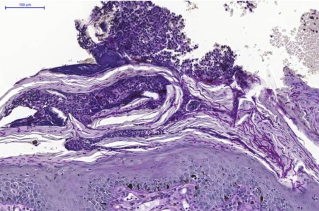
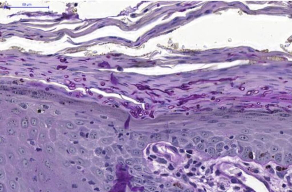
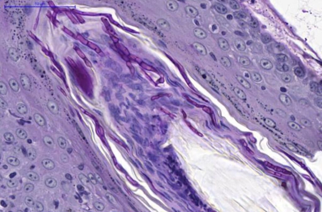
The samples exhibited a diffusely hyperplastic epidermis with mild acanthosis, occasional intracellular oedema, and multifocal moderate ortho- and parakeratotic hyperkeratosis. Multifocal superficial subcorneal pustules contained neutrophils, cellular debris, erythrocytes, eosinophilic material, and occasional acantholytic keratinocytes. Numerous 2-4 μm basophilic arthrospores and rare hyphae were identified within the keratin layers of the epidermis and hair follicles. Hair follicles were multifocally dilated, hyperkeratotic, and occasionally effaced by a marked neutrophilic and macrophagic inflammatory infiltrate (folliculitis and furunculosis). Periodic acid-Schiff (PAS) staining confirmed fungal septate hyphae within the epidermis and follicular keratin layers.
Interpretation
Based on the microscopic findings, a diagnosis of epidermal and follicular hyperplasia with hyperkeratosis, superficial pustules, numerous intralesional keratin-colonising fungal structures, and suppurative to pyogranulomatous folliculitis and furunculosis of the nasal skin was made.
Discussion
These microscopic changes were consistent with superficial pustular dermatophytosis, which was one of the clinical differential diagnoses in this case. PAS staining demonstrated numerous fungal elements within the keratin layers of the epidermis and hair follicles, findings compatible with keratin-colonising dermatophytosis. Culture of lesional tissue was recommended to identify the fungal species involved.
In dogs, dermatophytosis is most commonly caused by Microsporum canis, although other agents include M. gypseum, Trichophyton mentagrophytes, Epidermophyton floccosum, and T. rubrum. Lesions are typically localised to the face, pinnae, paws, and tail, and present as patches of alopecia with variable papules, pustules, scaling, and crusting. Breed predispositions have been reported, with T. mentagrophytes infections more frequent in Parson Russell Terriers, and M. canis infections in Yorkshire Terriers and Pekingese.
Dermatophytes, with rare exceptions, colonise only cornified structures, including hair shafts, the inner root sheath, the stratum corneum, and claws/nails and do not invade viable tissue.
The main differential diagnoses for this case include pemphigus foliaceus, superficial spreading pyoderma, and candidiasis. Pemphigus foliaceus typically demonstrates more pronounced acantholysis than is observed in most cases of superficial pustular dermatophytosis. Superficial spreading pyoderma is characterised by the presence of blue line formation, a feature absent in superficial pustular dermatophytosis. Pustular folliculitis, when present, is strongly suggestive of dermatophytosis, particularly in cats, and concomitant mural folliculitis is also a characteristic finding in this type of fungal infection. In all cases, however, demonstration of fungal elements is essential for a definitive diagnosis, and culture may be required to differentiate superficial pustular dermatophytosis from candidiasis.
References
1-Gross TL, Ihrke PJ, Walder EJ, Affolter VK. Skin Diseases of the Dog and Cat: Clinical and Histopathologic Diagnosis. 2nd ed. Oxford: Blackwell Science Ltd; 2005. p. 11–13. doi:10.1002/9780470752487.
2-Peters J, Scott DW, Erb HN, Miller WH Jr. Comparative analysis of canine dermatophytosis and superficial pemphigus for the prevalence of dermatophytes and acantholytic keratinocytes: a histopathological and clinical retrospective study. Vet Dermatol. 2007;18(4):234–40. doi:10.1111/j.1365-3164.2007.00599.x. PMID:17610488.
3-Moriello KA, Coyner K, Paterson S, Mignon B. Diagnosis and treatment of dermatophytosis in dogs and cats: Clinical consensus guidelines of the World Association for Veterinary Dermatology. Vet Dermatol. 2017;28(3):266–e68. doi:10.1111/vde.12440. PMID:28516493.