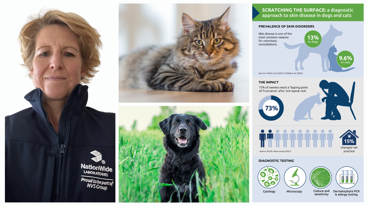
Scratching the surface: a diagnostic approach to skin disease in dogs and cats
Author: Stacey Newton BVSc FRCPath CertEM (Int Med) PhD MRCVS
Skin disease is one of the most common reasons for veterinary consultations. In the UK, skin disorders affect around 13% of the dog population under primary veterinary care,1 while in cats, the prevalence is 9.6%, after dental disease and obesity.2 Despite their frequency, skin conditions can be diagnostically challenging, with similar clinical presentations masking a wide range of underlying causes.
The impact of skin diseases extends beyond the animal, significantly affecting owners as well. Studies have shown that owners of dogs with chronic skin conditions experience heightened caregiver burden, leading to physical exhaustion, mental fatigue, social difficulties, family disagreements, and financial strain.3 This burden can strain the vet-client relationship, especially when treatment plans are complex or outcomes are not as expected. Notably, a survey revealed that 73% of clients reached a ‘tipping point of frustration’ after the third veterinary visit for dermatological issues. Of these, 15% discontinued using their veterinary practice altogether.4
Effective history-taking
A systematic approach is essential for accurate diagnosis and effective treatment. No dermatology diagnostic work-up is complete without a thorough history. Gathering detailed background information can help narrow the differential list, reduce the number of diagnostic procedures needed and help with interpreting results.5 Details such as age, breed, onset and progression of signs, seasonality, parasite control, contact with other animals, and previous treatment all help refine the differential list. A standardised dermatology history form can be useful in complex cases, saving time in consults and highlighting patterns that may otherwise be missed.
Beyond first impressions
A careful clinical examination is essential in any dermatology case, beginning with a general examination to identify systemic signs that might suggest endocrine or metabolic disease. This should be followed by a detailed dermatological examination, including inspection of the skin, ears and mucocutaneous junctions.
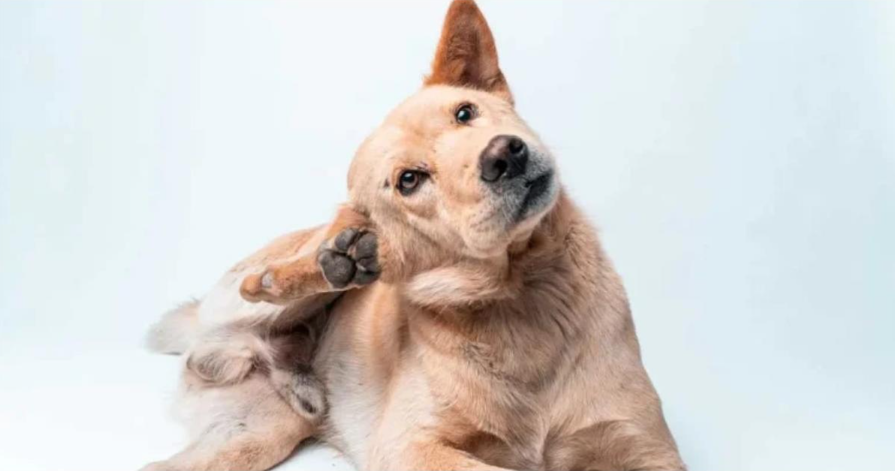
Note the distribution of lesions and whether they are primary (e.g. papules, pustules or macules) or secondary (e.g. alopecia, crusts, lichenification). The pattern and location of lesions can offer valuable diagnostic clues – such as lesions on the ear margins in sarcoptic mange or facial alopecia and crusts in feline dermatophytosis. With a comprehensive history and clinical examination, it is often possible to make a tentative diagnosis.5
Essential diagnostic techniques
Although some cases may initially respond to empirical treatment, identifying the underlying cause is often necessary, especially in chronic cases or those that fail to respond to trial treatment. Initial diagnostic tests include cytology, direct microscopy, and culture.
Cytology
Cytology remains a cornerstone of the diagnostic process. It is quick, minimally invasive and cost-effective and alongside histopathology, it provides immediate visual insights into pathological processes. Samples from skin lesions, ear canals or interdigital spaces can enable rapid identification of bacterial infection, yeast (particularly Malassezia spp,) and inflammatory cell types.
Common cytological techniques include:
Impression smears: For superficial skin lesions (scabs, ulcers, pustules or vesicles) identifying infectious organisms including Staphylococcusand Malassezia. Any scab material should be removed and a clean glass slide touched onto the surface of the lesion. Gently squeezing the skin may help with visualisation of Demodex mites.
Tape strips: Often used for identification of Malassezia, particularly where skin folds or the local anatomy prevents good contact between the slide and the skin surface.
Swabs: Smears made from swabs may be used for superficial skin lesions (pustules, vesicles or draining skin tracts) and are also the method of choice for ear cytology. Where necessary, the swab should be moistened with 0.9% NaCl. The smear should be prepared by gently rolling the swab across the slide; rubbing the swab across the slide is not advised as it will cause cell damage.
Skin scrapings: For identification of sarcoptic or demodectic mange mites, or to harvest cells from ulcerated skin lesions e.g. eosinophilic granuloma complex. To obtain deep scrapings, for detection of Demodex mites, the skin should be gently squeezed until capillary bleeding is noted.
Dermatophyte diagnosis
Microscopy of hair samples can yield additional information – spores indicative of dermatophyte infection, evidence of follicular dysplasia or dystrophy, or broken hair shafts indicative of self-trauma induced alopecia for example. However, when it comes to dermatophytes, direct microscopy of hair samples alone does not exclude dermatophytosis and dermatophyte culture or PCR may be indicated.
For many years, fungal culture has been considered the gold standard of dermatophyte diagnosis. However, it may take between 7 to 14 days for definitive results, during which time infection may progress or be passed to other animals or people. Dermatophyte PCR offers a rapid and reliable alternative, typically providing results within a few days. Quicker diagnosis facilitates prompt initiation of appropriate treatment and environmental management strategies. For this reason, PCR is increasingly preferred, especially in cases where rapid confirmation or exclusion of dermatophyte infection is necessary, such as outbreaks in multi-animal environments or where there is zoonotic risk. Similarly to fungal culture, PCR results should always be interpreted alongside clinical signs.
Culture and sensitivity testing
For bacterial infections, culture and sensitivity testing is particularly beneficial in the diagnosis and management of canine pyoderma. An ideal sample for culture is obtained by lancing a pustule with a sterile needle after preparing the surface aseptically to remove surface contaminants. Culture and sensitivity testing identifies the bacterial species involved and the antibiotics effective against this strain. This allows precise selection of an appropriate antibiotic treatment (if systemic use is required) and is never contraindicated.6 It is important to note that the antibiotic sensitivity data does not apply to topical use.7 Given recent concerns over methicillin-resistant Staphylococcus pseudintermedius (MRSP), culture and sensitivity testing is increasingly vital to prevent further antibiotic resistance.8
Identifying allergic skin disease
And finally, allergic skin disease represents a considerable proportion of chronic dermatological conditions in veterinary practice. These include atopic dermatitis, food hypersensitivities, and flea allergy dermatitis (FAD). Clinical signs typically include pruritus, erythema, and secondary lesions such as alopecia, crusts, and excoriations resulting from intense itching. An accurate diagnosis of allergic conditions depends heavily on a thorough history, noting the onset, progression, and seasonality of clinical signs, and any response to previous treatments or dietary changes.
Diagnostic options include serological allergy testing or intradermal testing for environmental allergens, as well as carefully conducted elimination diet trials for suspected food allergies. Effective allergy management often involves a combination of allergen avoidance, dietary modifications, immunotherapy, and targeted medical therapy to control clinical signs.
Given their complexity, effective management of dermatological conditions relies on accurate and timely diagnostics. A structured diagnostic approach helps improve treatment outcomes, reduce the likelihood of chronic or recurring skin conditions, and ultimately improve both animal welfare and client satisfaction.
References
- O’Neill, D.G. et al. (2021) Prevalence of commonly diagnosed disorders in UK dogs under primary veterinary care: results and applications. BMC Vet Res 17, 69 (2021). https://doi.org/10.1186/s12917-021-02775-3)
- O’Neill, D.G. et al. (2023) Commonly diagnosed disorders in domestic cats in the UK and their associations with sex and age. Journal of Feline Medicine and Surgery. 25(2). doi:10.1177/1098612X231155016
- Spitznagel, M.B. et al. (2019) Caregiver burden in the veterinary dermatology client: comparison to healthy controls and relationship to quality of life. Vet Dermatol, 30: 3-e2. https://doi.org/10.1111/vde.12696
- American College of Veterinary Dermatology survey of 300 clients (2021)
- Lundberg, A. et al. (2022) Diagnosing common skin conditions of dogs and cats in community medicine practice. TVP
- Tait, J. (2022) Pyoderma in the Dog. Today’s veterinary nurse.
- Allerton, F. and Nuttall, T. (2021) Antimicrobial use: importance of bacterial culture and susceptibility testing. In Practice, 43: 500-510. https://doi.org/10.1002/inpr.139
- Bajwa, J. (2016) Canine superficial pyoderma and therapeutic considerations. The Canadian veterinary journal. 57(2), 204–206
About the Author
Stacey Newton BVSc FRCPath CertEM (Int Med) PhD MRCVS
Stacey Newton is Head of Clinical Pathology at NationWide Laboratories and a veterinary pathologist with extensive expertise in diagnostic testing. After graduating from Bristol in 1993, she gained clinical experience when working as a locum in general practice for six months before having a permanent job in a mixed practice in Pontefract. In 1995 she started an internal medicine residency in equine medicine at the University of Liverpool-based mainly at Leahurst. She obtained her certificate in equine medicine (internal medicine) during that time. At the end of the residency, she went on to do a PhD in equine neurology, also based at the University of Liverpool. This was mainly based on headshaking in horses and working with Dr Knottenbelt. Part of the work was with Paul Eldridge a Neurosurgeon at The Walton Hospital, Liverpool. His speciality was Trigeminal neuralgia. The residency and PhD were funded by The home of Rest for Horses and Horseracing Betting Levy Board, respectively. The PhD was obtained in 2002. She then locumed for a variety of veterinary practices for six months before then joining Nationwide Laboratories which was then a privately-owned laboratory. She continued to work for the same laboratory as a clinical veterinary pathologist. She obtained her Diplomat of the Royal College of pathologists in 2008 and then went on to do become a Fellow of the Royal College of pathologists in 2010. She has various published papers during her time at university on equine medicine. She has also talked at the BEVA Congress and other venues providing CPD. Since then, she has been a key figure in clinical pathology, providing veterinary professionals with expert interpretation of laboratory results. Stacey’s deep understanding of diagnostic techniques offers valuable insights across a range of conditions, including dermatological disease, helping vets make informed clinical decisions with confidence.
Original publication: Veterinary Edge, issue 51, May 2025, pp 46-47