Mineral associated lymphadenopathy in a dog
Author: Karina Fresneda
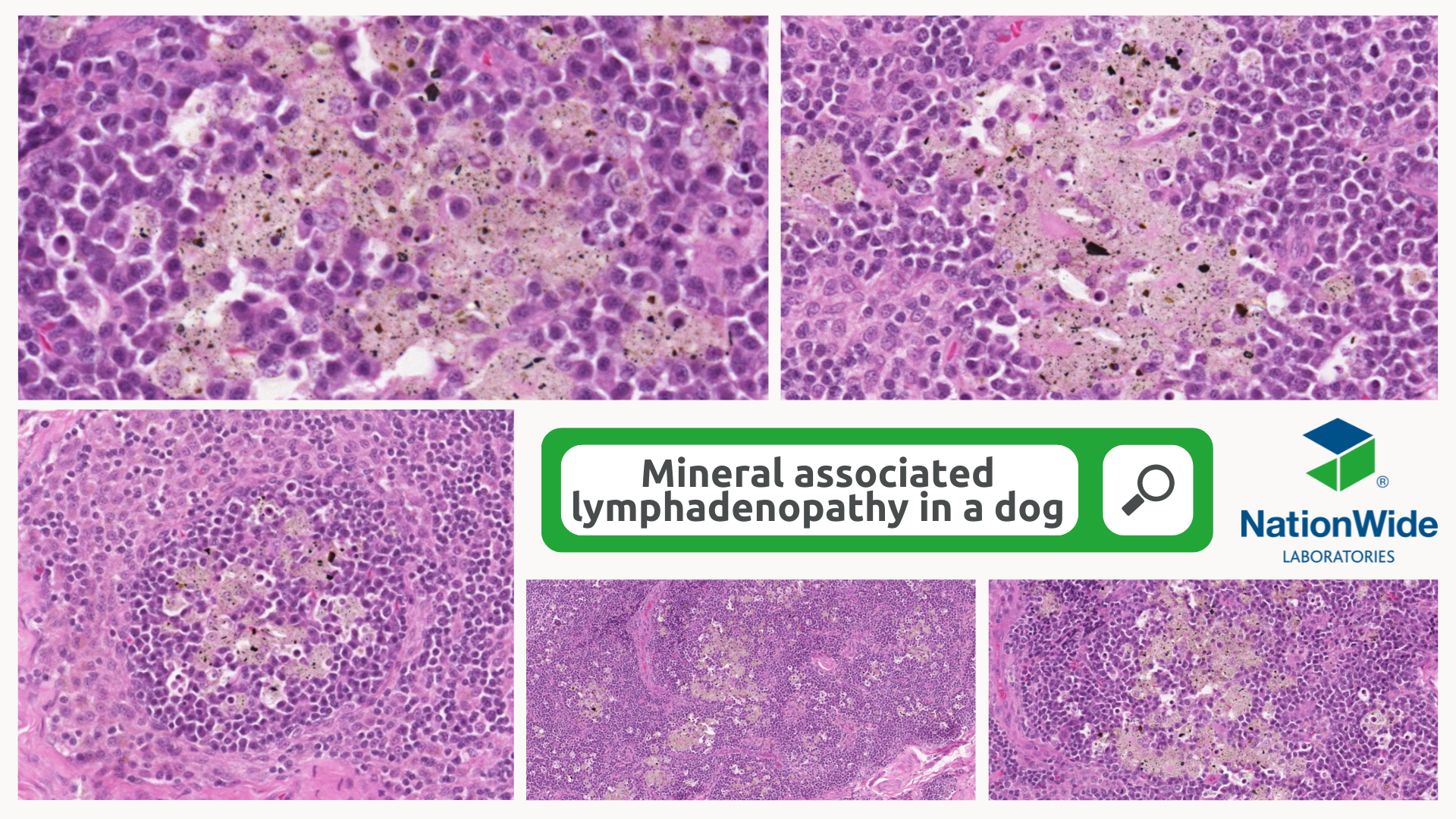
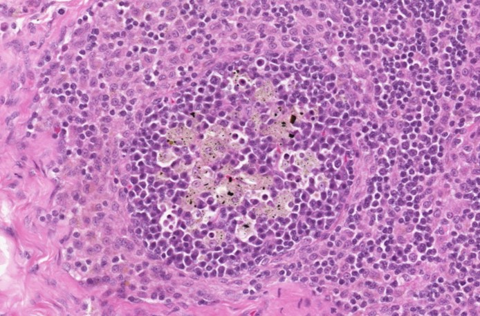
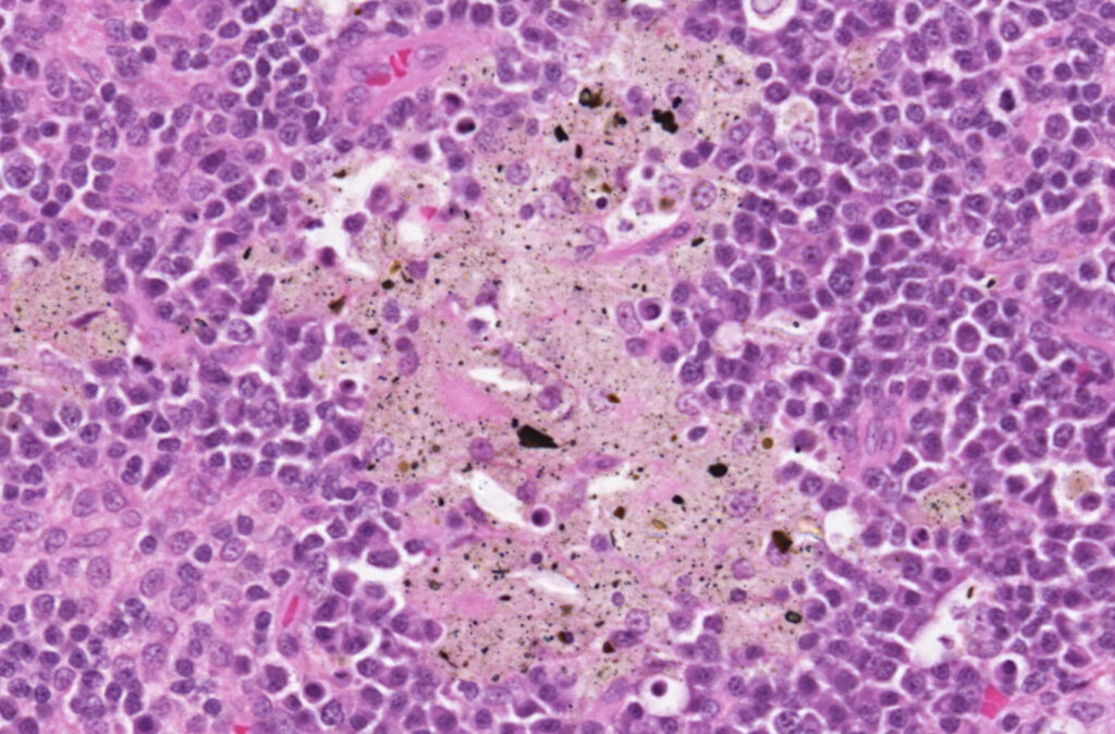
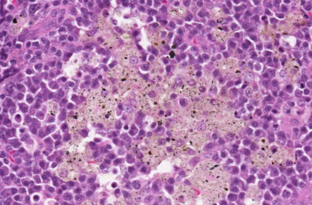
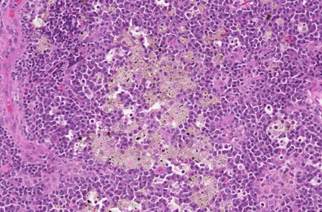
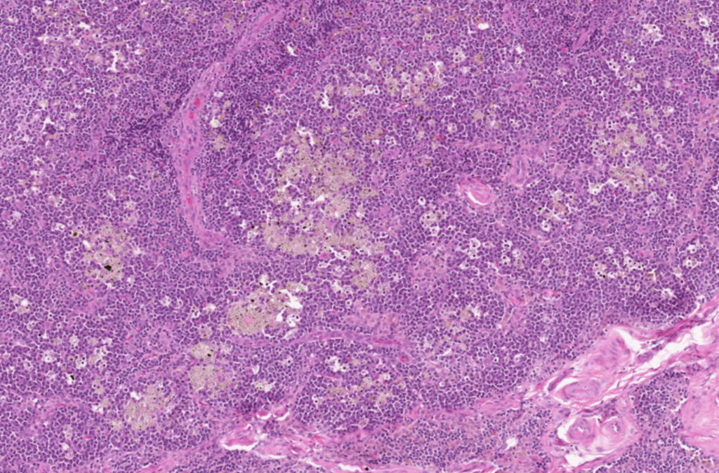
We received samples from prescapular and thoracic lymph nodes. Microscopically, the medullary sinuses were expanded by numerous plasma cells, small to intermediate lymphoid cells and macrophages, the latter were often laden with birefringent crystalline brown yellowish material. The diagnosis was mineral associated lymphadenopathy. This condition was first documented in the UK in the late 1980s, and it causes enlargement of lymph nodes. The pathogenesis is still unknown. The most common clinical signs in dogs are lymphadenopathy and non-specific signs such as pyrexia or inappetence. Histopathology is needed to confirm the diagnosis where the microscopic lesions are granulomatous lymphadenitis with accumulation of crystalline, mixed mineral material within the node, that is phagocytosed by macrophages. Electron microscopy of these nodes have shown that this mineral comprise a complex mixture of elements, such as aluminium, silicon, titanium or nickel, that are presumed to have an environmental origin; however, the source of them, portal of entry and mechanism of deposition within the peripheral lymph nodes has not yet been determined.
References:
Lesions Associated with Mineral Deposition in the Lymph Node and Lung of the Dog
M. J. Day, G. R. Pearson, V. M. Lucke, S. J. Lane, and R. S. J. Sparks https://journals.sagepub.com/doi/pdf/10.1177/030098589603300104
Differential Diagnosis of Lymphadenopathy. Michael J. Day. World Small Animal Veterinary Association World Congress Proceedings, 2004.