Granular cell tumour in an 8-year-old Pomeranian – case study

Credit: Dr Marvin Firth BVSc (Hons.), DipRCPath, DipFMS, AFHEA, MRSB, MRCVS
A mass that was attached to the underside of the tongue in an 8-year-old Pomeranian was noted on clinical examination by the primary veterinarian. This was surgically removed and sent for histopathological evaluation to NationWide Laboratories.
The tissue was composed of typical skeletal muscle and oral epithelium consistent with that of the tongue, yet focally there is also extensive ulceration. The underlying connective and muscular tissue is effaced and infiltrated by a densely cellular, unencapsulated, poorly demarcated mass. The mass is composed of sheets of densely packed, round to polygonal cells supported by a fibrovascular stroma (see Fig 1.). These neoplastic cells have variably distinct borders, often eccentrically placed round nuclei with abundant eosinophilic granular cytoplasm. There was mild to moderate pleomorphism of the cells and low mitotic activity. Unfortunately, the mass extends to the deep and lateral margins of the sections examined.
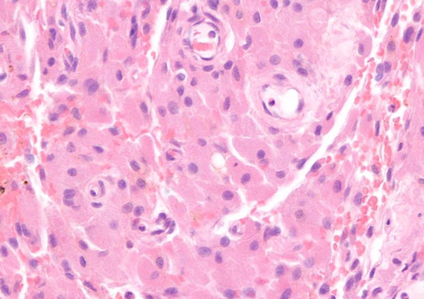
Fig 1. H&E, Granular cell tumour, dog. Neoplastic cells are densely packed and are round to polygonal with abundant eosinophilic granular cytoplasm.
Additional staining of the tissue with periodic acid Schiff (PAS), shows fine PAS positive granules within the cytoplasm of neoplastic cells (seen in Fig 2 and 3).
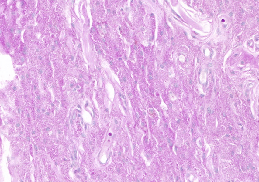
Fig 2. PAS, Granular cell tumour, dog. X40 Magnification. Densely cellular mass containing neoplastic cells with abundant finely PAS-positive granules.
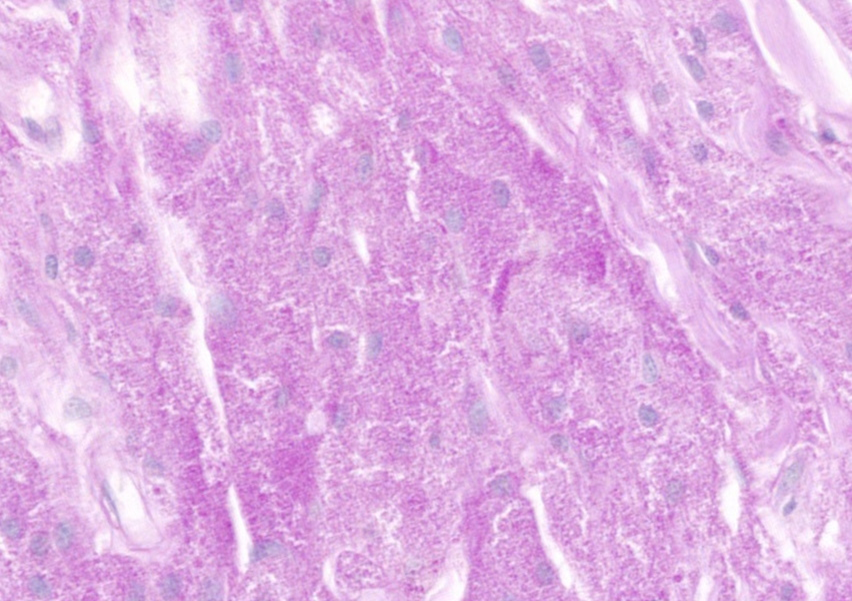
Fig 3. PAS, Granular cell tumour, dog. X60 magnification. Easily noted PAS positive granules within the cytoplasm of neoplastic cells.
The findings of the histopathology and additional stains confirmed the presence of a granular cell tumour of the tongue of this dog. These are fairly uncommon, yet benign tumours found in the skin and subcutis, oral cavity or even the central nervous system, as well as a common tumour in the lung of horses. The name is descriptive and does not imply a specific cell of origin or differentiation, which is, yet, unknown in these tumours. Immunohistochemistry adds little additional information as to the cell of origin, but these tumours tend to be well-demarcated, nonencapsulated and have variable amounts of collagenous stroma. Most granular cell tumours are benign and complete surgical excision is curative.
The clarity afforded by the 3DHISTECH system allows pathologists to see the greatest of detail at both a tissue and cytoplasmic level of the neoplastic cells. This enables us to make more informed and accurate diagnoses of tumours that may not have additional tests to confirm their histogenesis. In this case, we are also able to accurately report surgical margins with software tools that accurately measure and outline the neoplastic against the normal tissue. For more information about our histopathology and cytology services please visit our Histopathology and Cytology page.