Feline eosinophilic sclerosing fibroplasia

Credit: Karina Fresneda DVM DiplACVP
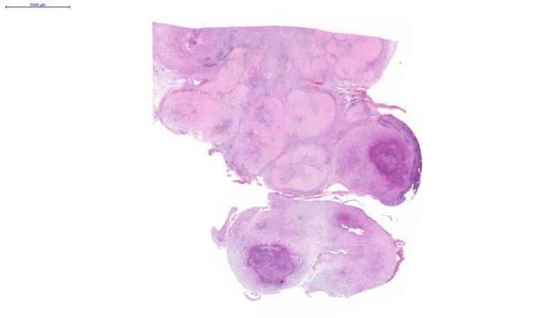
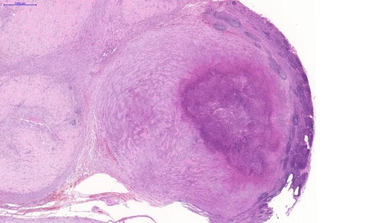
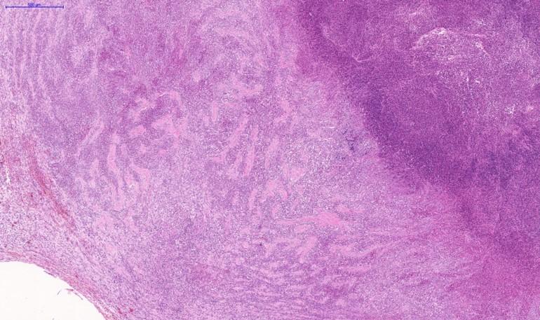
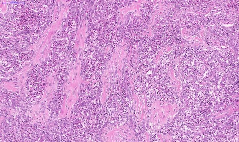
This is a section of ileum, where submucosa and tunica muscularis were almost entirely effaced by an extensive proliferation of irregular anastomosing trabeculae of collagen separated by numerous eosinophils and spindle cells, and fewer mast cells, histiocytes, neutrophils, lymphocytes and plasma cells. These findings are consistent with eosinophilic sclerosing fibroplasia. In this case was also observed in the colon.
Feline eosinophilic sclerosing fibroplasia is an unusual condition observed in cats. The aetiology is still unknown. Some cases were associated with bacteria, but antibiotic treatment was not effective. It has been hypothesized that genetically predisposed cats may develop FESF in response to the introduction of bacteria or other antigens into the intestinal wall. The disease was most often observed in middle-aged cats, that generally had a long history of vomiting and/or diarrhoea. Lesions are typically large, hard, non-painful, easily palpable and most commonly situated near the pylorus or ileocaecocolic junction; however, it has also been reported outside of the abdominal cavity. The lesion is often grossly, and might be misdiagnosed as osteosarcoma, due to the branching collagen trabeculae that resemble osteoid tissue, sclerosing mast cell tumour, due to the presence of eosinophils and mast cells, and sarcoma, because the reactive spindle cells shared many cytological features with sarcomatous mesenchymal cells. Cats treated with prednisone had a significantly longer survival time than those receiving other treatments.
References
Feline gastrointestinal eosinophilic sclerosing fibroplasia. L E Craig, E E Hardam, D M Hertzke, B Flatland, B W Rohrbach, R R Moore. Veterinary Pathology 2009 Jan;46(1):63-70.
A Case of Feline Gastrointestinal Eosinophilic Sclerosing Fibroplasia Associated with Phycomycetes. Grau-Roma, I.Galindo-Cardiel, M.Isidoro-Ayza, M.Fernández, N.Majó. Journal of Comparative Pathology. Volume 151, Issue 4, November 2014, Pages 318-321
Feline gastrointestinal eosinophilic sclerosing fibroplasia: 13 cases and review of an emerging clinical entity. Michael Linton, Judith S Nimmo, Jacqueline M Norris, Richard Churcher, Sophia Haynes, Agnieszka Zoltowska, Sunishka Hughes, Naomi S Lessels, Miranda Wright, Richard Malik. Journal of Feline Med Surg. 2015 May;17(5):392-404.
Ultrasonographic and clinicopathological features of feline gastrointestinal eosinophilic sclerosing fibroplasia in four cats. Andrea Weissman, Dominique Penninck, Cynthia Webster, Silke Hecht, John Keating, Linden E Craig. J Feline Med Surg. 2013 Feb;15(2):148-54.
A case of feline gastrointestinal eosinophilic sclerosing fibroplasia mimicking metastatic neoplasia. J S Munday, A W Martinez, M Soo. N Z Vet J, 2014 Nov;62(6):356-60.
Feline eosinophilic sclerosing fibroplasia – a characteristic inflammatory response in sites beyond the gastrointestinal tract: case report and proposed nomenclature. Bianca Zampieri, Molly E Church, Koranda Walsh, Elizabeth M Lennon. JFMS Open Rep. 2022 Aug 17;8(2):
A case of an intramural, cavitated feline gastrointestinal eosinophilic sclerosing fibroplasia of the cranial abdomen in a domestic longhair cat. Gordon A Davidson, Samantha S Taylor, Melanie J Dobromylskyj, Francesco Gemignani, Helen Renfrew. JFMS Open Rep. 2021 Feb 23;7(1)