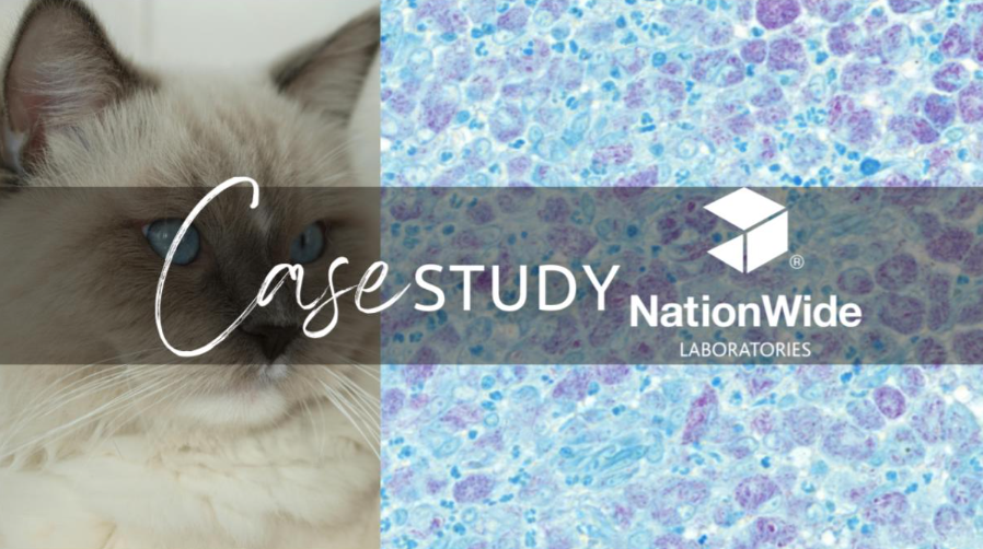
Digital cytology: possible Cryptococcosis in a 10-year-old domestic shorthair cat
Credit: Dr. Stacey A Newton BVSc FRCPath CertEM (Int Med) PhD MRCVS
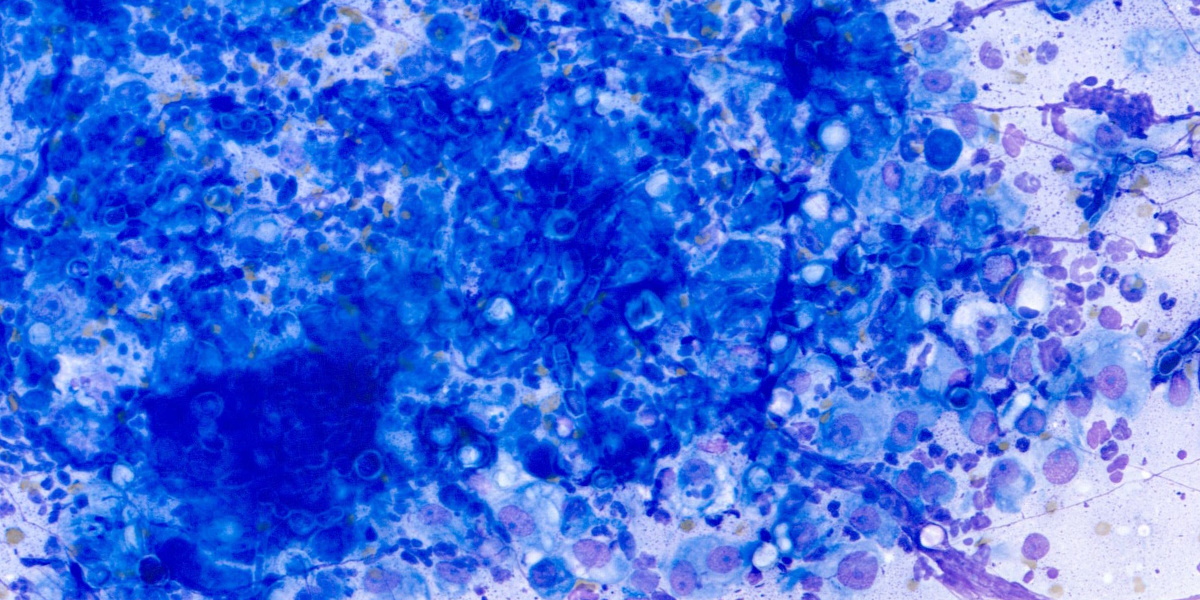
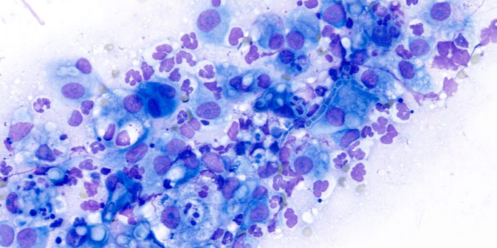
A 10-year-old domestic shorthair male cat presented with a recurrent nasal mass, which was initially excised surgically last year. The clinical pathology team at NationWide Laboratories performed a cytological examination of the mass using digital pathology images, revealing marked pyogranulomatous inflammation and suspicious yeast elements that were likely to be Cryptococcus. The presence of round to oval (11-12 um diameter) yeast structures with internal basophilic stippling and a thin clear external halo were observed both extracellularly and within macrophages. There were also frequent pseudohyphae observed in some areas. This looked consistent with a diagnosis of Cryptococcus infection. The digital images allowed for high-resolution visualisation of the fungal elements; however, further confirmation would require culture or PCR testing to definitively identify the species and determine the specific genotype involved.
The diagnosis of Cryptococcus typically involves confirmation through culture or PCR testing, which is crucial for determining the species and genotype involved. The presence of fungal elements in the cytology sample suggests that the infection is likely caused by the Cryptococcus neoformans or Cryptococcus gattii species (other species e.g. C. albidus and C. magnus are rare), which is a common cause of fungal infections in cats. The diagnosis was further supported by the presence of fungal-related histopathological changes in the biopsy sample.
Treatment for Cryptococcus typically involves a prolonged course of antifungal therapy, with Amphotericin B, ketoconazole, fluconazole, and itraconazole being commonly used. Surgical excision of any nodules may also be a valuable adjunct treatment to enhance treatment outcomes and prevent recurrence. In this case, the cat was treated with a combination of antifungal therapy and surgical excision of the nasal mass.
Further information
Cryptococcosis is considered the most common systemic fungal disease in cats. It is not a zoonotic disease, and infected cats are not contagious to humans and other animals. The disease is typically caused by the Cryptococcus neoformans-Cryptococcus gattii species complex and is acquired from contaminated environments.
The infection can manifest in various clinical forms, including nasal (most common), central nervous system, cutaneous, and systemic forms.
Diagnosis can be confirmed through diagnostic imaging, antigen detection, cytology, biopsy samples, and culture or PCR testing.
Treatment typically involves a prolonged course of antifungal therapy, with surgical excision of any nodules as an adjunct treatment.
CNS and systemic forms may require symptomatic treatment.
Prognosis is favourable in most cases if diagnosed early and treatment compliance is good.
Prevention. Free-roaming cats in rural areas are potentially more exposed to Cryptococcus, even though urban cats can be contaminated through pigeon guano. According to ecology, the presence of avian guanos, particularly pigeon droppings, and some decaying vegetation substrates, such as Eucalyptus leaves, may be considered risk factors.
Conclusion
This case demonstrates the benefits of integrating digital pathology technology into veterinary diagnostic workflows. The use of advanced digital imaging technology enabled rapid and accurate identification of fungal elements, allowing for prompt diagnosis and disease management. The collaboration between the practice team and NationWide Laboratories’ experts ultimately led to improved treatment outcomes for this feline patient.
Reference
Cryptococcosis in cats guidelines, first published in the J Feline Med Surg 2013, 15: 611-618 by Maria Grazia Pennisi et al. The present guidelines used here were updated by Maria Grazia Pennisi.