Dermal canine haemangioma
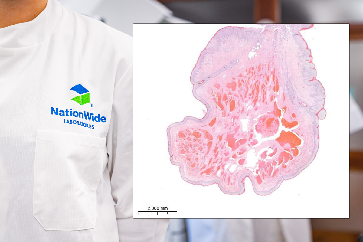
This is a skin mass from a 9-year-old crossbreed canine. This is a densely cellular, 7.5mm diameter, well-demarcated, unencapsulated, multinodular, pedunculated mass within the dermis of haired skin. It consists of vascular spaces of various calibre, which predominantly contain erythrocytes, but also some contain fibrin or pale eosinophilic material. These spaces are bordered by plump neoplastic endothelial cells and supported by variably abundant fibrocollagenous stroma that have rare small accumulations of lymphocytes and plasma cells. The proliferating endothelial cells have oval nuclei with finely stippled chromatin and indistinct nucleoli. There is moderate eosinophilic, fibrillar cytoplasm and indistinct cell borders. There is mild anisocytosis and mild anisokaryosis with typically less than 1 mitoses per high power fields (2.37mm2). Subepithelial dermal regions contain mild to moderate lymphoplasmacytic infiltrates with increased numbers of neutrophils within the dermis of the stalk. There are clear borders of haired skin in this tissue section.
In the dog, haemangiomas are typically benign, solitary, deep dermal tumours, whereas haemangiosarcomas are often present as a disseminated malignancy involving the spleen, heart, lung, liver, soft tissues of the trunk and extremities (Cooley et al., 1997). Accurate and clear digital imaging assist pathologists in determining whether a mass is benign or malignant by being able to assess the neoplastic cell population, in this case the fine endothelial cells lining the vascular channels. In dogs, cutaneous haemangiomas are common as compared to primary canine cutaneous haemangiosarcomas. Haemangiomas occur more frequently in younger dogs.