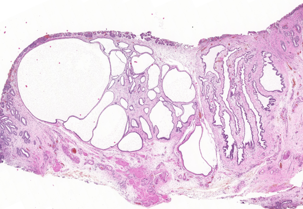
Cystitis Glandularis in a 13-year-old female cat

Credit: Karina Fresneda DVM DiplACVP
Cystitis is a clinically significant disease that affects various species, with a higher incidence in females, likely linked to their shorter urethra. This condition can manifest as either acute or chronic. Chronic cystitis takes on diverse forms based on the pattern and type of inflammatory response, including diffuse, follicular, and polypoid variants.
Chronic polypoid cystitis is characterized by the presence of polypoid masses, particularly common in dogs but may occur in any species. These masses likely develop as an inflammatory and hyperplastic response to chronic irritation, often stemming from persistent bacterial urinary tract infections (UTIs) and/or uroliths.
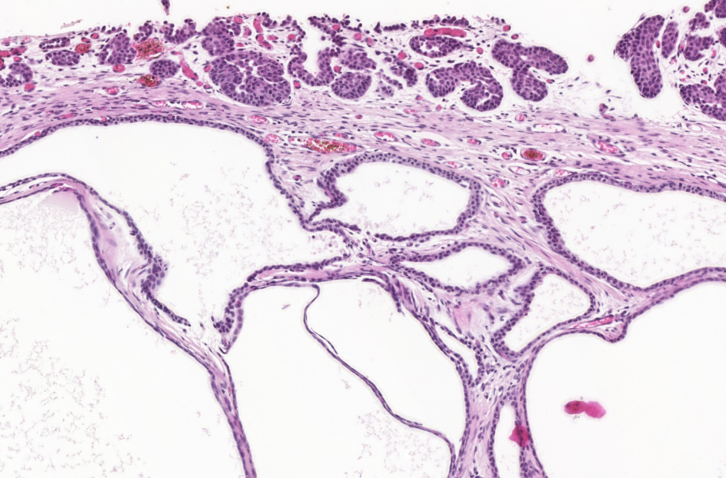
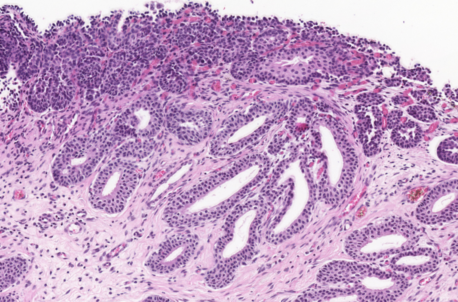
The polyps found in the bladder mucosa consist of a core of proliferative connective tissue covered by surface epithelium. This surface epithelium can form nests of hyperplastic urothelial cells in the lamina propria, known as Brunn’s nests, or undergo metaplasia to a mucus-secreting, glandular epithelial type, leading to a condition called CYSTITIS GLANDULARIS. These resulting polypoid masses may have a broad-based or pedunculated structure, occurring most frequently in the cranioventral bladder wall. Chronic haematuria, often unresponsive to antimicrobial therapy, is a common observation in cases of chronic polypoid cystitis. This bleeding is often associated with the breakdown of polyps, potentially promoting urine retention and bacterial introduction, thus establishing a cyclic pattern of infection, proliferation, and inflammation.
Histologically, our case had in lamina propria, occasionally extending from mucosa, multiple hyperplastic tortuous glands that were often cystic dilated. They were lined by one to multiple layers of uniform cuboidal or columnar epithelial cells, occasionally surrounded by a few layers of urothelial cells. They had no evident atypia. The lamina propria where they were immersed contained a few plasma cells, lymphocytes, neutrophils and rare histiocytes.
References
Cystitis glandularis in a cat. Amalia Agut, Juana D Carrillo, Marta Soler, Juan D Garcia, Eliseo Belda, Miguel A Gomez and Antonio Bernabe. Journal of Feline Medicine and Surgery. Volume 16, Issue 4, April 2014, Pages 363-365.
Cystitis Cystica, Cystitis Glandularis, and Brunn’s Nests in a Feline Urinary Bladder. J. F. Zachary. Veterinary pathology Journal. Volume 18, Issue 1.
Pathology of domestic animals. Volume 2. Jubb, Kennedy & Palmer’s. Sixth edition. 2016. Pages 459-461
Pathologic basis of veterinary disease. James F. Zachary. Seventh edition. 2022. Page 762