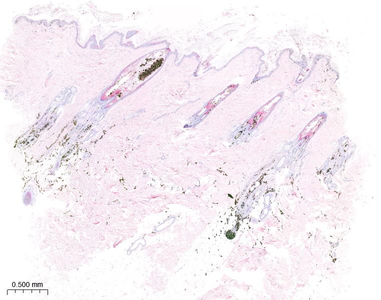
Colour dilution alopecia in a 3-year-old male Doberman

Here is a case from a 3-year-old male Doberman that was exhibiting generalised dry dermatitis and alopecia over the trunk and had been non-responsive to conventional, medical treatment. A series of biopsies were examined digitally here at NationWide Laboratories which showed mild changes within the epidermis including irregularly spaced, hyperpigmented basal epithelial cells and some pigmentary incontinence. Within the dermis, there was very noticeable follicular dysplasia and dense clumps of melanin pigment within the hair bulbs and follicles and even in the hair shafts, including enlarged melanomacrophages surrounding the base of the hair follicles. Overall, within the sections, the hair follicles are atrophied. There were minimal inflammatory cells throughout the sections. These histological features are consistent with chronic pigmentary intrafollicular and intraepidermal melanin clumping and pigmentary incontinence that is consistent with colour dilute/colour mutant alopecia or black hair follicular dysplasia and are observed in colour dilute dogs (e.g., blue or fawn colours). The follicular dysplasia often leads to poor hair coat quality and variably alopecia, such as that reported in this case. Hair loss is usually most severe on the trunk, especially dorsally. Secondary bacterial infection can sometimes be seen. This form of colour dilution alopecia is commonly seen in the Doberman Pinscher. As such, this is a classic case and the use of digital pathology allows us to both accurately diagnose the condition, whilst effectively sharing the key histological findings to our trainee pathologists and students. The ability to pan the whole slide image also allows us to quickly make judgements of the most important pathology and hone our attention to these changes.


