Amyloid-producing odontogenic tumour (APOT) in a 5-year-old Maine-Coon cross cat
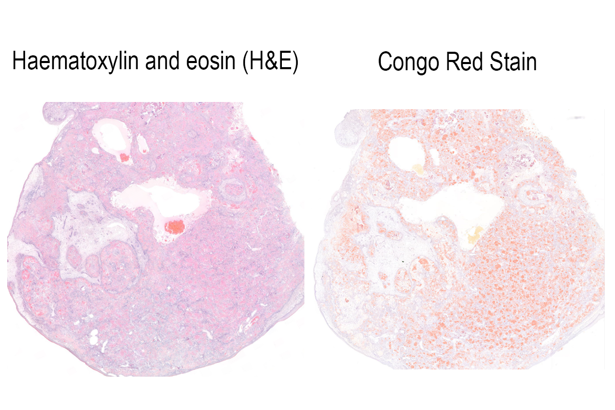
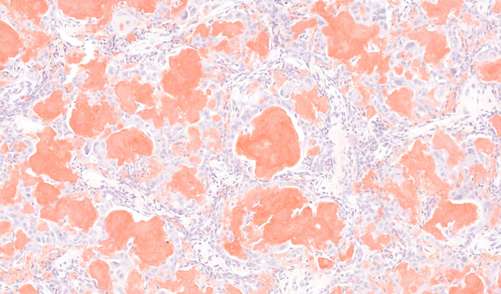
This is a piece of tissue taken as a biopsy from the mouth of a 5-year-old Maine-Coon cross cat. It was a hard piece of tissue for our technicians to process and when we examine this histologically, we can see that the submucosa is expanded and effaced by a densely cellular, poorly demarcated, and locally invasive proliferation of epithelial cells exhibiting cord, ribbon and island arrangements. Within the centre of many of the larger islands are circular to globular aggregates of brightly eosinophilic, smudged to glassy, acellular, amorphous material which was presumed to be amyloid.
A special stain, Congo Red (right-hand slide), highlights that this material is in fact, amyloid produced by the tumour. Digital pathology allows us to compare, side by side the tissue as examined with the routine haematoxylin and eosin, against special stains and immunohistochemistry. This allows us as pathologists to examine and capture the very nature of pathology changes allowing us to make accurate diagnoses. Here, we have an amyloid-producing odontogenic tumour (APOT).
These are rare tumours of the oral cavity and have been reported in dogs, cats, a goat, horses, rabbits, moose, Bengal tigers and prairie dogs!
They comprise approximately 1-4% of odontogenic neoplasms in the dog and have more recently been reported in the facial skin of cats.