NationWide Laboratories: Cutaneous plasmacytoma in a 7-year-old female Labrador

Author: Stacey Newton BVSc FRCPath CertEM (Int Med) PhD MRCVS
Case presentation
A 7-year-old entire female Labrador was presented with a 2 cm round firm haired dermal mass on the left-hand side rostral lip. The duration of the mass was not indicated. Further investigation with fine-needle aspirates for cytology were performed.
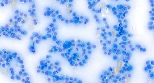
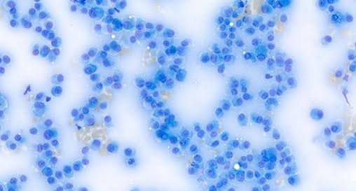
Cytology
This demonstrated a predominant population of round to polygonal shaped cells which had moderate anisocytosis and anisokaryosis. These looked round cell in origin. They had round to oval nuclei with stippled chromatin, single or multiple prominent nucleoli and moderate to sometimes large amounts of variably finely granular basophilic cytoplasm. Frequent cells showed varying amounts of irregularly shaped and sized nonstaining globular structures within the cytoplasm. Occasional cells had a small amount of coarse blue granular material within the cytoplasm. Most had an eccentric position to the nucleus. Occasional cells had macro nucleoli. Binucleate and multinucleate cells were frequently observed. There was an occasional clear zone to one side of the nucleus in some cells. A small amount of eosinophilic amorphous material was seen among the cells.
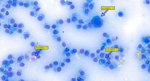
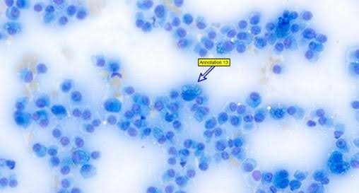
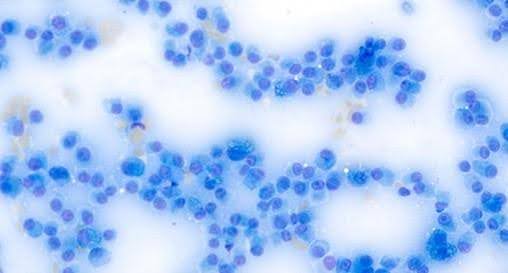
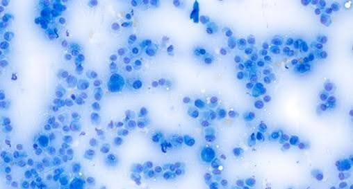
Interpretation
Round cell tumour most compatible with a plasmacytoma.
Further investigation
The mass was excised approximately 2 weeks later. A piece of skin and tissue measuring 19 x 16 mm with a raised ulcerated mass measuring 7 mm diameter was submitted for histology.
Histology
Sections were prepared and demonstrated a raised ulcerated nodular mass at the centre of a piece of haired skin. This was composed of sheets of atypical neoplastic nongranular round cells. The cells had large oval nuclei often with prominent nucleoli and coarsely clumped chromatin patterns. They had abundant cytoplasm. The occasional multinucleate cells and rare cells resembling antibody laden Mott cells were observed. Mitotic figures were 2-3 per 10 high-power fields (x400). The nodule was discrete but not encapsulated.
No pictures as done on glass.
Interpretation
Ulcerated cutaneous plasmacytoma.
Comment
Plasmacytomas are common cutaneous neoplasms of the dog, accounting for approximately 2% of all canine skin tumours. These usually occur in older dogs with an average age of approximately 10 years. They are usually solitary small slightly raised dermal nodules. These are often covered by alopecic skin which is occasionally ulcerated. The most common sites are pinnae and digits however other parts of the face, ear canal as well as oral cavity can be affected. Despite the cytological atypia these are typically benign and usually respond to complete surgical excision. Occasional recurrence following surgical excision has been reported usually where complete excision is difficult. Rare cases of metastasis to distant skin sites as well as peripheral blood involvement have been reported. Multiple forms have been reported in the absence of multiple myeloma. Note multiple myeloma is usually associated with other vague systemic clinical signs, monoclonal gammopathy or hypercalcaemia.
Recent appear more susceptible to developing plasma cell tumours include Airedale Terriers, Cocker Spaniels, Kerry Blue Terriers, Scottish Terriers, and Standard Poodles. Multiple myeloma is seen more often in German Shepherds, Labradors and Golden Retrievers.
Plasmacytomas are rarely reported in the cat. When they occur, they have been reported to be found at several sites including paws, thorax, face (lip, chin), neck, shoulder, tail, metatarsals and nose. Less is known about the clinical behaviour in cats due to the low numbers of reports. For masses localised to the skin and/or regional lymph nodes surgical excision has been reported to usually result in long-term resolution. Early progression to systemic disease has been reported in a few cases.