Correlating digital cytopathology and neoplastic cavitary effusion in a 12-year-old cat

Author: Paulina Woźniak
An entire female 12-year-old domestic shorthair cat was presented to the veterinary clinic, where clinical evaluation diagnosed an abdominal mass, suspected of pancreatic origin and the evidence of abundant peritoneal fluid. Fine-needle aspirates of the mass and peritoneal fluid sample were submitted to a UK veterinary diagnostic laboratory. The slides were prepared using cytospin technique and assessed by digital cytopathology. The samples were found to have cytological features consistent with an epithelial neoplasm, highly suggestive of adenocarcinoma.
The predominant population in samples from the mass consisted of moderately to markedly pleomorphic epithelial cells, with round nuclei, finely stippled nuclear chromatin, prominent nucleoli and moderate to abundant basophilic cytoplasm. The cells were usually distributed in clusters and occasionally presented pseudo-acinar arrangements. The slides from peritoneal effusion revealed similar population of the cells, admixed with inflammatory component.
Cavitary effusion parameters
Appearance of fluid – milky pale yellow
Total nucleated Cell Count – 11.3 x 10^9/l
Red Cell Count – <0.01 x 10^12/l
Total Protein – 43 g/l
Albumin – 24 g/l
Triglycerides – 6.62 mmol/l
Cytological findings
The predominant population in the submitted samples consisted of moderately to markedly pleomorphic epithelial cells, with round nuclei, finely stippled nuclear chromatin, prominent nucleoli and moderate to abundant basophilic cytoplasm. The cells were usually distributed in clusters and occasionally presented in pseudo-acinar arrangements. The slides from the peritoneal effusion revealed a similar population of cells, admixed with a predominantly neutrophilic inflammatory component.
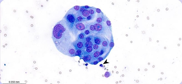
Figure 1. Abdominal mass. Photomicrograph presenting clusters of markedly pleomorphic epithelial cells. The size of the cells can be compared to a neutrophil (arrowhead). Leishman stain, x50 objective.
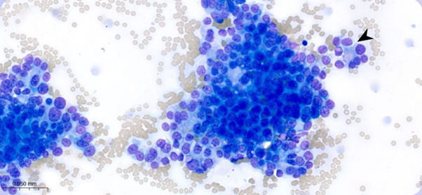
Figure 2. Abdominal mass. Photomicrograph presenting a larger cluster of relatively uniform epithelial cells showing vague acinar type arrangements, suggesting secretory origin (arrowhead). Leishman stain, x50 objective.
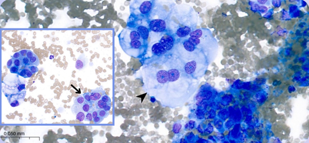
Figure 3. Abdominal mass. Photomicrographs presenting clusters of markedly pleomorphic epithelial cells. Binucleation (arrowhead) and vague acinar structures (arrow) are noted. Leishman stain, x50 objective.
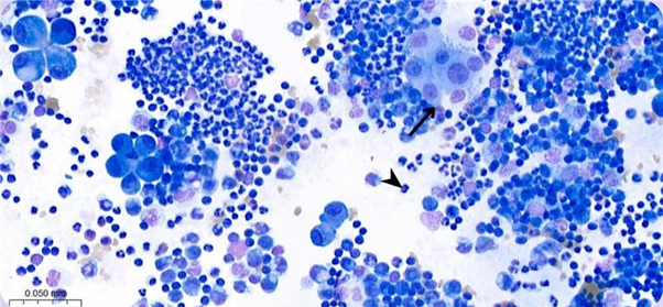
Figure 4. Peripheral effect. Photomicrograph of neoplastic, inflammatory effect presenting clusters of pleomorphic cells (arrow). The size of the cells can be compared to a neutrophil (arrowhead). Leishman stain, x50 objective.
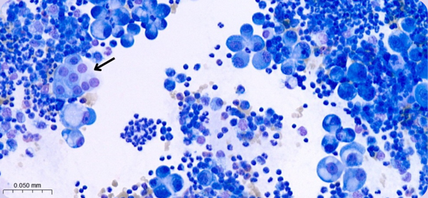
Figure 5. Peripheral effusion. Similar to Figure 4, with one group of cells resembling epithelial appearance (arrow). Leishman stain, x50 objective.
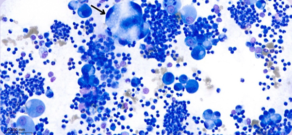
Figure 6. Peripheral effusion. Photomicrograph of a large pleomorphic cluster of cells (arrow). Leishman stain, x50 objective.
Discussion
Pancreatic adenocarcinoma is uncommon in dogs and cats; however, it is the most common tumour of the exocrine pancreas. There are no breed or sex predispositions. The clinical signs may include vomiting, abdominal pain, inappetence, weight loss. The most common sites of metastasis are liver, peritoneum and local lymph nodes. The median survival time of cats with abdominal effusion was 30 days compared with 97 days for cats with no effusion. Cytology is generally considered an easy, safe, relatively non-invasive diagnostic tool for diagnosing neoplastic processes. However, there is one case report of suspected needle tract seeding of pancreatic adenocarcinoma. In presented case, the cat was euthanized 33 days after first symptoms occurred due to rapid deterioration. Other epithelial neoplastic processes, such as metastatic cholangiocellular carcinoma or renal carcinoma could be mentioned as differential diagnoses but are considered less likely given the localization of the mass and clinical findings. Further diagnostic tests, such as histology of the mass or immunocytochemistry could provide more information regarding the exact origin of the cells. Our findings reinforce the fact that pancreatic carcinoma should be rated highly in the differential diagnosis in mature adult and senior cats with abdominal masses, ascites and/or jaundice.
References
Linderman, Michael & Brodsky, E & de Lorimier, Louis-Philippe & Clifford, C & Post, Gerald. (2012). Felineexocrine pancreatic carcinoma: A retrospective study of 34 cases. Veterinary and comparative oncology. 11.10.1111/j.1476-5829.2012.00320.x. 1.
Julie Allen, Chapter 8 – Pancreas (exocrine and endocrine), Editors: Rose E. Raskin, Denny J. Meyer, Katie M.Boes, Canine and Feline Cytopathology (Fourth Edition), W.B. Saunders, 2023, p. 322-338, ISBN 9780323683685. 2.
Jegatheeson S, Dandrieux JR, Cannon CM. Suspected pancreatic carcinoma needle tract seeding in a cat. JFMSOpen Rep. 2020 Jun 1;6(1):2055116920918161. doi: 10.1177/2055116920918161. PMID: 32537237; PMCID:PMC7268146.

