Equine biochemistry and haematology analysis
Author: Stacey Newton
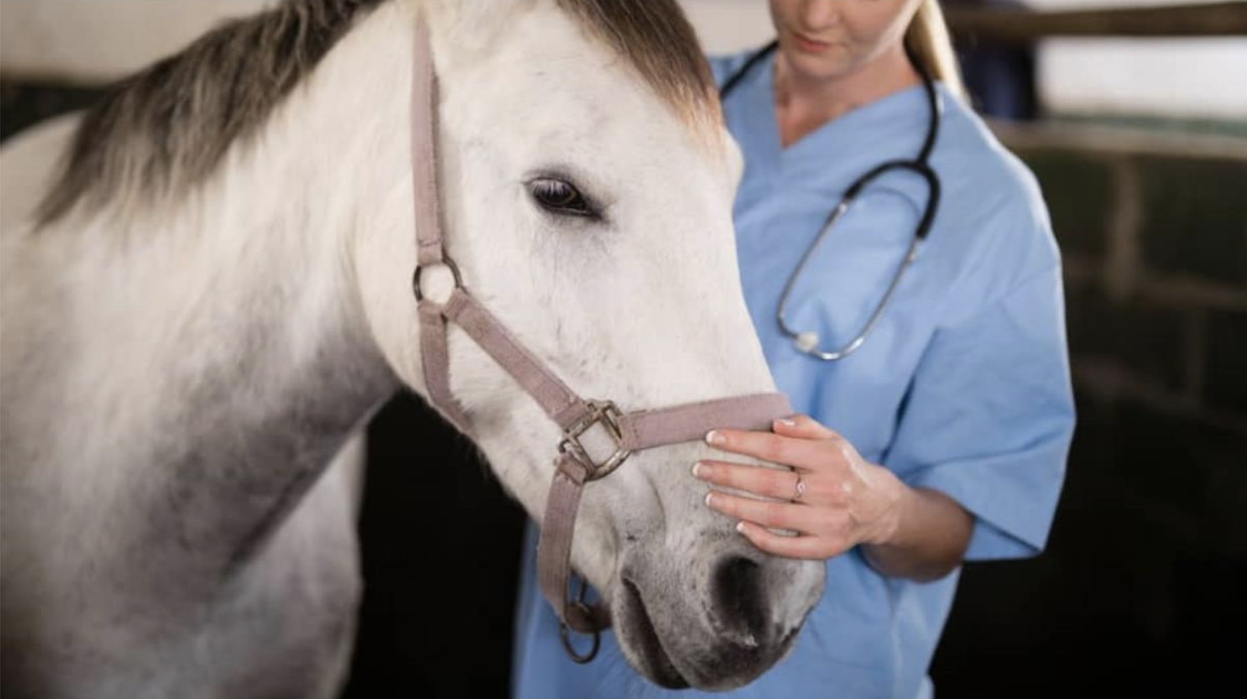
The use of haematology and biochemistry is vital in diagnosing many conditions in our equine patients. The timings of taking samples, the tests we request and the interpretation of results are skills we develop as practising clinicians. However, it is not uncommon to be reaching for the phone when our patient’s results come back to discuss the case with the pathologist.
We are extremely fortunate to have experienced pathologists working in our diagnostic labs in the UK. Here, the author shares her expertise, answering questions asked by and relevant to first opinion vets, shedding light on what we are seeing and how best to interpret results.
Q: Acute inflammatory markers are useful to evaluate the kinetics of inflammation, but are there any other reasons to use them?
A: The acute phase proteins include globulins, fibrinogen, high serum amyloid A (SAA), iron and albumin. As well as giving us useful information on inflammation, they can also provide insights to other conditions.
Fibrinogen can be used as a marker of disseminated intravascular coagulation (DIC) in horses, as it is low in many cases of DIC. In these cases, it should be considered alongside D-dimers, platelets, prothrombin time (PT) and activated partial thromboplastin time.
Iron (Fe) is reduced with inflammation but can also be used as a marker of Fe deficiency anaemia alongside a reduced mean corpuscular volume (MCV; microcytic). But be aware that it is not specific on its own as it may also decrease with haemolysis and chronic anaemia.
Albumin is a negative acute phase protein and when significantly decreased, suggests other causes such as protein loss – for example, protein-losing enteropathy, protein-losing nephropathy and, although rare, hepatic insufficiency.
Q: What is the importance of D-dimers in inflammation in horses, both young and adults? Also, can I combine fibrinogen, SAA and D-dimers?
A: D-dimers are not specific for inflammation as they are part of the coagulation cascade and then fibrinolysis. They are a product of fibrinolysis, and increases are seen in any age of patient.
A D-dimer is a specific degradation fragment of cross-linked fibrin and a high plasma D-dimer is an indicator of intravascular fibrin formation and plasmin-mediated fibrinolysis.
The measurement of plasma D-dimer concentration is useful to aid in the diagnosis of systemic thrombosis, including pulmonary thromboembolism and DIC.
D-dimers can be combined with fibrinogen if suspicion of DIC exists. Fibrinogen and SAA are acute phase proteins that are increased with inflammation.
However, SAA has a much shorter half-life than fibrinogen, so combining the two can be used to determine the course of inflammation and whether it is ongoing or resolving.
Q: Is there any evidence that higher levels of SAA indicate an infectious process rather than just an inflammatory process?
A: We do not have a particular level of SAA that is more likely to indicate an infectious process, but bacterial disease is more likely to be present in many cases (Long and Nolen-Walston, 2020).
Elevations in SAA can increase with inflammation in the absence of infection and are not specific for a certain disease process.
Therefore, elevations in SAA should be evaluated in conjunction with physical examination findings and the results of other diagnostic tests.
Q: Why is polychromasia not seen in horses and why, then, can we use mean corpuscular volume and mean corpuscular haemoglobin concentration?
A: Horses don’t release their red blood cells from the bone marrow until they are more mature than other species, and as such we don’t see polychromasia. By the stage they do release their red blood cells they will have also lost their RNA, which gives the blue tinge to polychromatic cells with normal haematoxylin and eosin (H and E) stains.
However, the red blood cells are larger in size, meaning the MCV is greater. This can be of use in some cases to suggest regeneration, depending on the time of sampling in relation to the incidence of haemorrhage or haemolysis. If mean corpuscular haemoglobin concentration (MCHC) is increased, which in normal circumstances is not physically possible, MCHC suggests intravascular haemolysis, indicating that free haemoglobin is present, as well as that measured in the red blood cells.
The article continues beyond this point.
Read the full article https://www.vettimes.com/clinical/equine/equine-biochemistry-and-haematology-analysis
Original publication: Vet Times (2024), Volume 54, Issue 44, Pages 18-20
About the author
Stacey Newton, BVSc FRCPath CertEM (Int Med) PhD MRCVS is Head Veterinary Clinical Pathologist at NationWide Laboratories. She qualified from the University of Bristol in 1993 and completed a residency and PhD in equine medicine and neurology at the University of Liverpool. A Fellow of the Royal College of Pathologists, Stacey has published research in equine medicine and regularly contributes to CPD events, including BEVA Congress. More information on our equine services is available here https://nwlabs.co.uk/equine-service-list