NationWide Laboratories: Cytologic-histologic Correlation in Uncommon Neoplasms

Author: Dante Meza Ruiz, DVM, M.Ed., DVSc, Dipl. ACVP/ASVCP
A 10-year-old intact male Staffordshire Bull Terrier was presented to the veterinary clinic with a history of an enlarged popliteal lymph node on the right hind limb. A fine-needle aspiration (FNA) was performed, and the sample was submitted to NationWide Laboratories in the UK for evaluation.
Cytologic findings
The cytologic evaluation revealed a prominent lipid-rich background. The cellular population was predominantly arranged in cohesive clusters, with fewer cells observed individually. Most cells were round to polygonal, occasionally exhibiting spindle-shaped morphology. The cytoplasm was abundant and grey-blue with variably distinct borders, often containing conspicuous foamy vacuoles.
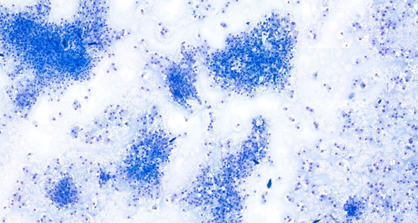
Figure 1
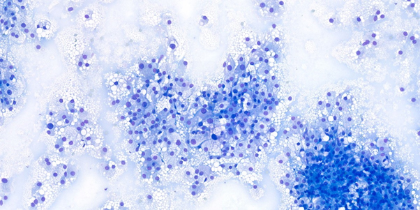
Figure 2
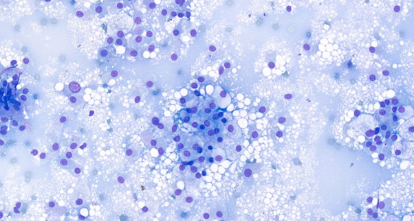
Figure 3
Figures 1-3: Photomicrographs of direct smears from a fine-needle aspirate of a mass labelled “popliteal lymph node” in a dog, stained with Leishman stain. (1) Numerous highly cohesive and individualised pleomorphic cells observed, x20 objective. (2) Cells exhibiting variably sized clear vacuoles and occasional binucleation, x40 objective. (3) Large round to polygonal cells with pale grey, occasionally granular cytoplasm, often showing clear vacuolation, along with round nuclei and prominent, single, round nucleoli, x60 objective.
Interpretation
A diagnosis of a malignant neoplasm was established, with no evidence of lymphoid tissue identified in the aspirate. Differential diagnoses included liposarcoma, clear cell adnexal carcinoma, sebaceous carcinoma, and balloon cell melanoma.
Additional test
An excisional biopsy and histopathologic evaluation were performed on the mass. A 2 cm elliptical, subcutaneous beige mass was submitted from the popliteal region. Histologically, the mass consisted of a discrete, unencapsulated subcutaneous lesion adhered to adjacent muscle. It was composed of cohesive sheets of large, round to polyhedral cells with abundant pale eosinophilic cytoplasm that was slightly granular to markedly lipidized. The nuclei were round to ovoid, hyperchromatic, and contained distinct nucleoli, with moderate anisonucleosis. Mitotic activity was rare, with fewer than one mitosis per high-power field. The tumor exhibited a small amount of fibrovascular stroma. The excision was extensively marginal, with portions of attached muscle included in the sample. The final diagnosis was subcutaneous liposarcoma, localized to the popliteal region.
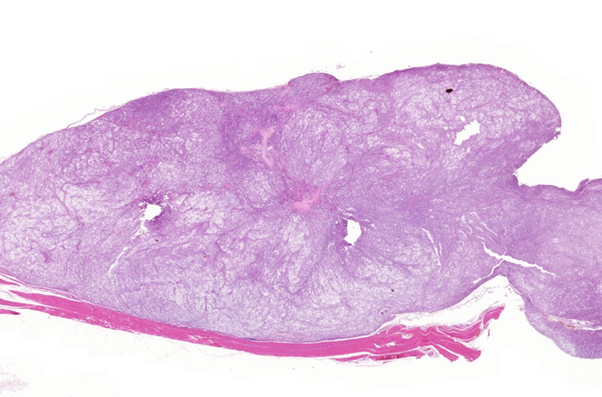
Figure 4
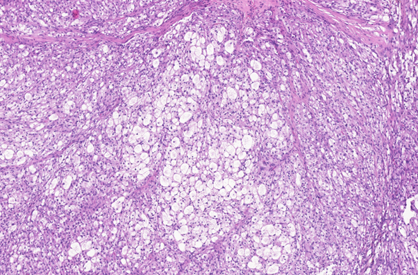
Figure 5
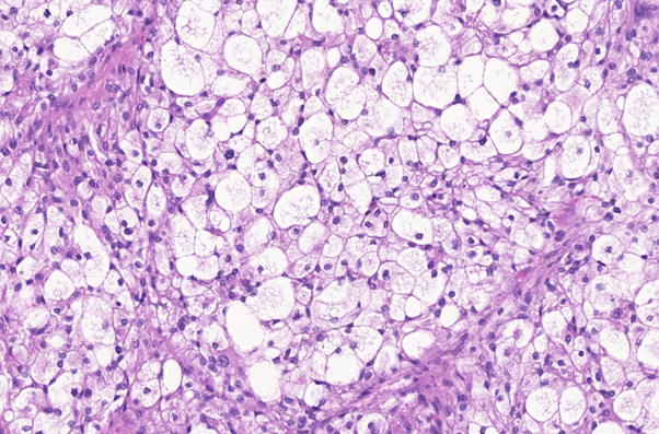
Figure 6
Figures 4-6: Photomicrographs of tissue sections from a subcutaneous mass in a dog. H&E stain. (4) Discrete, unencapsulated subcutaneous mass attached to adjacent muscle, x10 objective. (5) Clusters of tightly organized, large cells with round to polygonal shapes, x20 objective. (6) Cells with abundant pale eosinophilic cytoplasm, slightly granular to markedly lipidized, with round to ovoid, hyperchromatic nuclei, x40 objective.
Discussion
Liposarcomas are rare malignant tumours in dogs and cats that originate from lipoblasts, the precursor cells for fat tissue. These tumours are typically firm, poorly circumscribed, and attached to underlying tissues, often arising in subcutaneous locations. Due to their variable cytologic appearance and rarity, diagnosing liposarcomas through cytology can be challenging, often requiring histopathologic examination for a definitive diagnosis1.
Cytological analysis of this tumour was challenging due to the presence of features indicative of multiple tumour types. The cell morphology, which included polygonal to round shapes and areas of clustering, suggested carcinoma, while areas with indistinct cell borders and “naked nuclei” were more characteristic of a neuroendocrine tumour. Additionally, regions with loosely arranged, wispy cells were suggestive of sarcoma. The prominent lipid-rich background, along with intracellular vacuolation, led to consideration of liposarcoma, sebaceous carcinoma, and balloon cell melanoma. Ultimately, the diverse cytologic features and signs of malignancy pointed to a diagnosis of malignant neoplasia. Ultimately, the combination of various malignant characteristics and the cytologic features associated with different tumour types led to a diagnosis of malignant neoplasia.
On histopathology, well-differentiated liposarcomas are characterized by polygonal neoplastic cells with abundant, variably vacuolated cytoplasm embedded in a sparse collagenous stroma. Key features such as karyomegaly and nuclear pleomorphism distinguish these tumours from lipomas2. Special stains, such as Oil Red O, can aid in the diagnosis by highlighting lipid droplets in neoplastic cells. However, it is important to note that Oil Red O staining requires unprocessed tissue samples, as the lipid droplets are removed during the tissue processing phase involving alcohol fixation3.
Treatment of liposarcomas typically involves wide surgical excision to achieve clear margins, though additional therapies such as radiation and hyperthermia may be used to help control recurrence. Despite aggressive treatment, the prognosis is generally guarded, as liposarcomas have a high recurrence rate, though they rarely metastasize2.
References
1. Hendrick, M. J., Mahaffey, E. A., Moore, F. M., Vos, J. H., & Walder, E. J. (1998). Histological Classification of Mesenchymal Tumors of Skin and Soft Tissues of Domestic Animals. 2nd Series, Vol 2. Washington, DC: Armed Forces Institute of Pathology.
2. Raskin, R. E., Meyer, D. J., & Boes, K. M. (2022). Canine and Feline Cytopathology: A Color Atlas and Interpretation Guide (4th ed.). St. Louis, MO: Elsevier.
3. Masserdotti, C., Bonfanti, U., De Lorenzi, D., & Ottolini, N. (2006). Use of Oil Red O stain in the cytologic diagnosis of canine liposarcoma. Veterinary Clinical Pathology, 35(1), 37-41.


